. 2017 Sep;22(5):751-766.
doi: 10.1007/s12192-017-0802-0. Epub 2017 Jul 17.

A pyrrole-based natural small molecule mitigates HSP90 expression in MDA-MB-231 cells and inhibits tumor angiogenesis in mice by inactivating HSF-1

Affiliations

A pyrrole-based natural small molecule mitigates HSP90 expression in MDA-MB-231 cells and inhibits tumor angiogenesis in mice by inactivating HSF-1

K C Rashmi et al. Cell Stress Chaperones. 2017 Sep.

Abstract

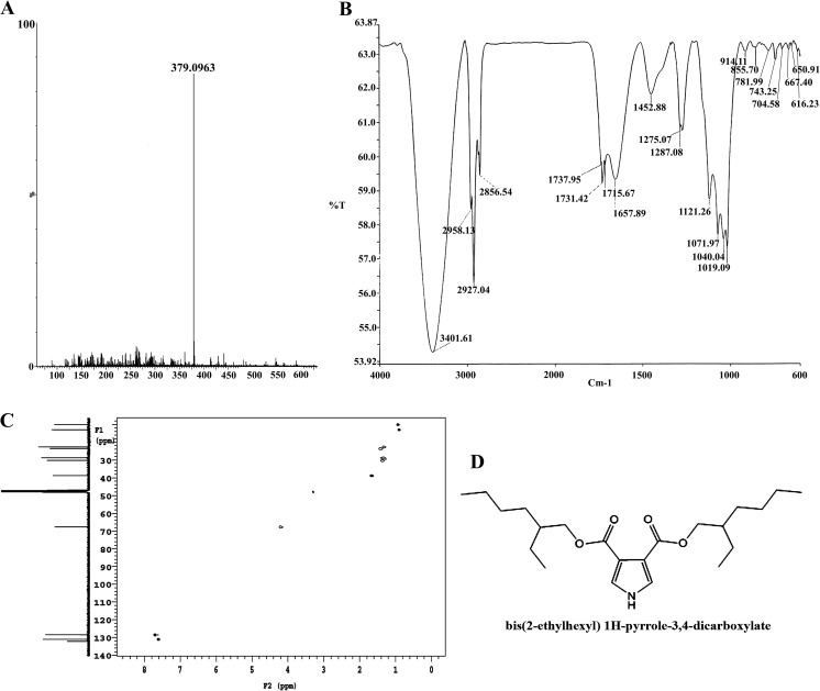
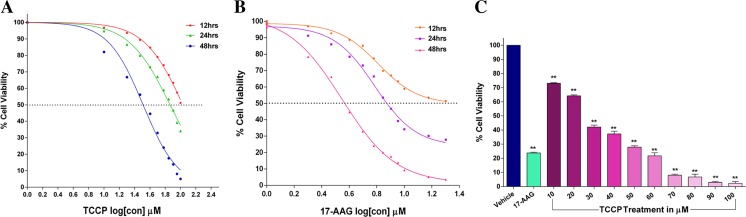
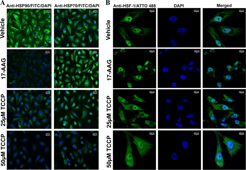
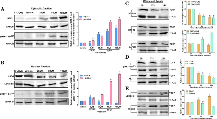
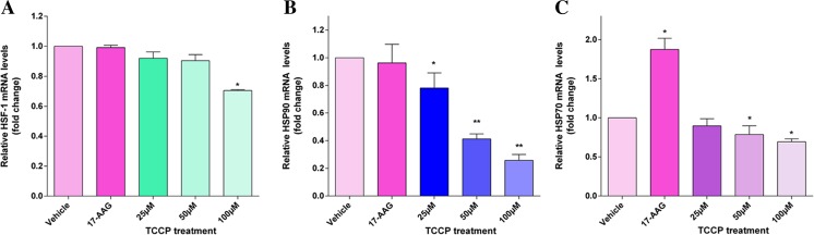
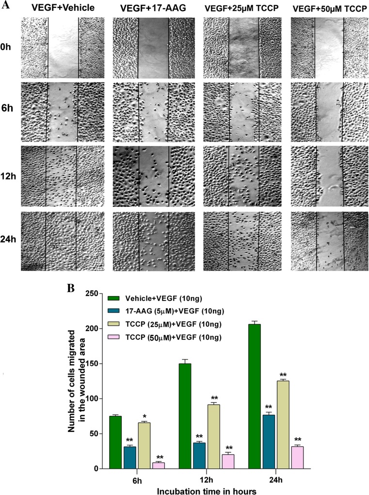
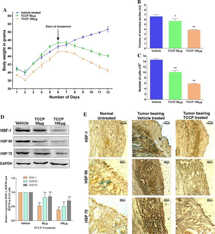
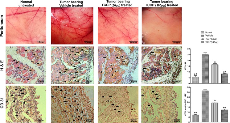
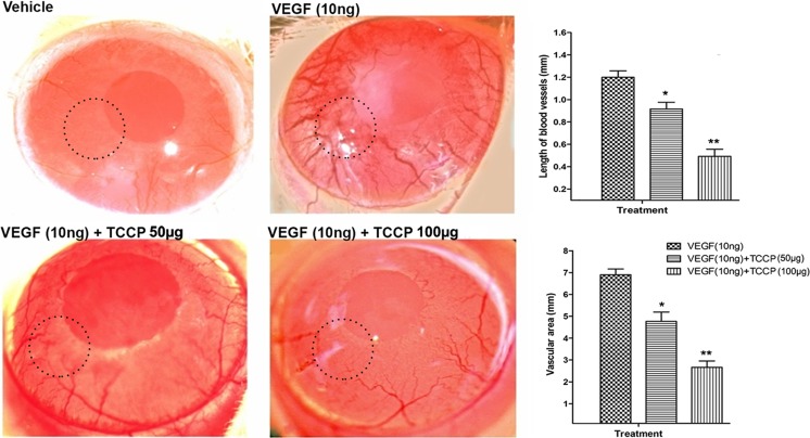
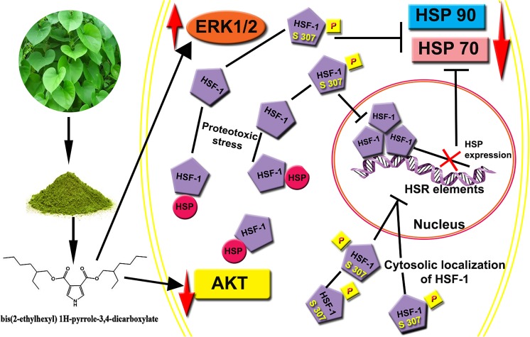
Heat shock proteins (HSPs), molecular chaperones, are crucial for the cancer cells to facilitate proper functioning of various oncoproteins involved in cell survival, proliferation, migration, and tumor angiogenesis. Tumor cells are said to be "addicted" to HSPs. HSPs are overexpressed in many cancers due to upregulation of transcription factor Heat-shock factor 1 (HSF-1), the multifaceted master regulator of heat shock response. Therefore, pharmacological targeting of HSPs via HSF-1 is an effective strategy to treat malignant cancers like triple negative breast cancer. In the current study, we evaluated the efficacy of a pyrrole derivative [bis(2-ethylhexyl)1H-pyrrole-3,4-dicarboxylate], TCCP, purified from leaves of Tinospora cordifolia for its ability to suppress heat shock response and angiogenesis using MDA-MB-231 cells and the murine mammary carcinoma: Ehrlich ascites tumor model. HSP90 was downregulated by TCCP by inactivation of HSF-1 resulting in inhibition of tumor cell proliferation, VEGF-induced cell migration, and concomitant decrease in tumor burden and neo-angiogenesis in vivo. The mechanism of suppression of HSPs involves inactivation of PI3K/Akt and phosphorylation on serine 307 of HSF-1 by the activation of ERK1. HSF-1 and HSP90 and 70 localization and expression were ascertained by immunolocalization, immunoblotting, and qPCR experiments. The anti-angiogenic effect of TCCP was studied in vivo in tumor-bearing mice and ex vivo using rat corneal micro-pocket assay. All the results thus corroborate the logic behind inactivating HSF-1 using TCCP as an alternative approach for cancer therapy.

Keywords: HSF-1; HSP; MDA-MB-231; NMR; Pyrrole; Tinospora cordifolia.


Figures

Fig. 1
Fig. 1
Characterization. a LS-MS spectrum. b Infrared spectrum. c 2D [13C-1H] heteronuclear single quantum correlation (HSQC) NMR spectrum. d Molecular structure of TCCP
Fig. 2
Fig. 2
Cytotoxicity and metabolic response of TCCP or 17-AAG on MDA-MB-231 cells. a In vitro cytotoxicity and IC50 assessment of TCCP treatments by trypan blue dye exclusion assay. Cells (3 × 104) were treated with either TCCP (10 to 100 μM/mL) or b 17-AAG (1 to 20 μM/mL) for 12, 24, and 48 h; washed with PBS; trypsinized stained; and counted in a hemocytometer. Data are presented as the mean ± SEM of three independent experiments. c Metabolic response to increasing concentrations of TCCP treatments assessed by MTT assay. Cells (3 × 104) were treated with TCCP, 17-AAG, or vehicle for 24 h and incubated with MTT solution, and the formazan crystals dissolved in DMSO and read at 570 nm. Data are presented as the mean ± SEM of three independent experiments. **p < 0.01, *p < 0.05
Fig. 3
Fig. 3
TCCP suppresses HSP expression and initiates cytosolic translocation of HSF-1. a Representative images of cellular localization of HSP90 and HSP70 post TCCP or 17-AAG treatments assessed by immunolocalization studies. Cells (5 × 104) were grown on coverslips, treated with TCCP or 17-AAG or vehicle, and incubated for 24 h and processed as described in the “Material and methods” section and photographed. b Representative images of immunolocalization of HSF-1 transcription factor post TCCP or 17-AAG treatment by confocal microscopy. Indirect immunofluorescent labeling of HSPs and HSF-1 proteins was by Atto 488-tagged secondary antibody and nuclei counterstained using DAPI
Fig. 4
Fig. 4
TCCP downregulates HSP90 and HSP70 through repression of HSF-1 activation. Dose response of TCCP induced repression of the HSF-1 transcription factor assessed by immunoblot analysis. a Cytosolic protein fraction. b Nuclear protein fraction. Confluent MDA-MB-231 cells in 100-mm petri dishes were treated with 17-AAG, vehicle, or TCCP for 24 h. The cytosolic and nuclear fractions were prepared following a modified method of Dignam fractionation, and clarified lysates (80 μg) were electrophoresed on 12% SDS-PAGE and transferred onto PVDF membranes. After blocking with 5% BSA/TBS, membranes were probed with anti-HSF-1 or anti-pHSF-1Ser307 separately for cytosolic and nuclear fractions. c 17-AAG and TCCP induced modulation of HSP90 and HSP70 expression and d AKT and e ERK phosphorylation at the indicated time points assessed by immunoblotting using whole cell RIPA buffer lysates. HRP-tagged secondary antibody was used for probing. GAPDH was used as an internal loading control for cytosolic or whole cell lysate blots and Lamin B1 for nuclear fraction blots and developed using the ECL method. Data are presented as the mean ± SEM of three independent experiments; **p < 0.01, *p < 0.05, and ns is p > 0.05
Fig. 5
Fig. 5
Relative mRNA transcript levels of HSF-1, HSP90, and HSP70, post TCCP or 17-AAG treatments assessed by qPCR. a TCCP or 17-AAG treatments have negligible influence on gene expression of HSF-1. b HSP90 gene expression is significantly decreased by TCCP but unaltered by 17-AAG. c HSP70 gene expression is marginally decreased by TCCP but drastically increased by 17-AAG. Total RNA was isolated from vehicle-, 17-AAG-, and TCCP-treated cells, and equal amounts of RNA were converted into cDNA and the mRNA levels of HSF-1, HSP90, and HSP70 were quantified by qPCR and normalized to GAPDH expression. Data are presented as the mean ± SEM of three independent experiments. *p < 0.05 significant and **p < 0.01 significantly different from the control
Fig. 6
Fig. 6
Suppression of cell migration by TCCP assessed by wound healing assay. a Confluent MDA-MB-231 cells in a six-well plate were serum starved overnight and treated with mitomycin c, and a scratch was made on the cell monolayer. Cell debris was washed, and the cells were cultured in basal medium containing VEGF along with indicated concentration of TCCP, 17-AAG, or vehicle. The wound closure was photographed at indicated time intervals. Images are representative of at least 10 independent locations that were monitored over the length of each wound. b Quantification of the cells involved in wound closure.*p < 0.01 = non-significant and **p < 0.01 considered significantly different from the control. Values are expressed as mean ± SEM of three replicate analyses
Fig. 7
Fig. 7
TCCP inhibits EAT cell proliferation and downregulates HSF-1, HSP90, and HSP70 in vivo. a Reduction in the body weight of EAT-bearing mice. b Cell number and c secreted ascites volume in untreated and treated groups. EAT-bearing mice were injected (i.p) with TCCP or vehicle from the 6th day of transplantation till the 12th day. The mice were sacrificed, cells were pelleted and counted, and volume of ascites was noted. d Immunoblot analysis and quantification of HSF-1, HSP90, and HSP70 protein expression in control and TCCP-treated EAT cells; cells were harvested from mice post treatment, and whole cell RIPA buffer lysates were prepared and immunoblotted for HSF-1, HSP90, and HSP70 proteins. GAPDH was used as an internal loading control. e Representative photographs of immunohistochemical staining for HSF-1, HSP90, and HSP70 proteins in the peritoneal section of tumor-bearing control, TCCP-treated, and normal mice. Post treatment, mice were sacrificed and the peritoneum was dissected and 5-μm microtome sections were stained using IHC methodology for HSF-1, HSP90, and HSP70 using respective antibodies and HRP-tagged secondary antibody and photographed using a bright-field microscope. Values are expressed as mean ± SEM of three replicate analyses; **p < 0.01,*p < 0.05, and ns is p > 0.05
Fig. 8
Fig. 8
TCCP mediated inhibition of peritoneal angiogenesis in vivo. Representative photographs of vascularization changes in the peritoneal wall upon TCCP treatment. Peritoneum sections of normal and EAT-bearing mice with indicated treatments. Lane 1—normal mouse (non-tumor-bearing), lane 2—control (tumor-bearing) treated with vehicle (0.1% DMSO in PBS), lane 3—tumor-bearing, treated with 50 μg TCCP, and lane 4—tumor-bearing, treated with 100 μg TCCP. The number of newly formed capillaries was assessed by the peritoneum pictures. MVD assessed by hematoxylin and eosin staining of peritoneum sections. CD31-positive staining was assessed by PECAM-1 immunohistochemical staining of peritoneum sections. Arrows indicate the presence of endothelial cell lining and infiltrating tumor cells in the peritoneum of the mouse. Values are expressed as mean ± SEM of three replicate analyses.*p < 0.05 significant and **p < 0.01 significantly different from the control
Fig. 9
Fig. 9
The corneal neovascularization inhibited by TCCP. Representative photographs of neovascularization observed in rat corneas: hydron polymer with vehicle (0.1% DMSO in PBS) negative control, hydron polymer + VEGF (10 ng) positive control, and hydron polymer + VEGF + TCCP (50 or 100 μg) were tests. Histograms showing length of blood vessels and vascular area in control and treated corneas both assessed by ImageJ software. Details of the experiments are described in the “Material and methods” section. After 7 days of incubation, the corneas were photographed at ×40 magnification through a stereo zoom microscope. Values are expressed as mean ± SEM of three replicate analyses.*p < 0.05 significant and **p < 0.01 considered extremely significantly different from the VEGF-treated positive control
Fig. 10
Fig. 10
The graphical representation of mode of action of TCCP-induced HSP downregulation in MDA-MB-231 cells

Similar articles

Cited by

References

    1. Adhvaryu MR, Reddy N, Parabia MH. Anti-tumor activity of four ayurvedic herbs in dalton lymphoma ascites bearing mice and their short-term in vitro cytotoxicity on DLA-cell-line. African Journal of Traditional, Complementary and Alternative Medicines. 2008;5(4):409–418. doi: 10.4314/ajtcam.v5i4.31297. - DOI - PMC - PubMed
    1. Bhardwaj V, Gumber D, Abbot V, Dhiman S, Sharma P. Pyrrole: a resourceful small molecule in key medicinal hetero-aromatics. RSC Adv. 2015;5(20):15233–15266. doi: 10.1039/C4RA15710A. - DOI
    1. Brewster AM, Chavez-MacGregor M, Brown P. Epidemiology, biology, and treatment of triple-negative breast cancer in women of African ancestry. The Lancet Oncology. 2014;15(13):e625–e634. doi: 10.1016/S1470-2045(14)70364-X. - DOI - PMC - PubMed
    1. Bruns AF, Yuldasheva N, Latham AM, Bao L, Pellet-Many C, Frankel P, Stephen SL, Howell GJ, Wheatcroft SB, Kearney MT. A heat-shock protein axis regulates VEGFR2 proteolysis, blood vessel development and repair. PLoS One. 2012;7(11):e48539. doi: 10.1371/journal.pone.0048539. - DOI - PMC - PubMed
    1. Carmeliet P, Jain RK. Molecular mechanisms and clinical applications of angiogenesis. Nature. 2011;473(7347):298–307. doi: 10.1038/nature10144. - DOI - PMC - PubMed

MeSH terms

Substances