Review
. 2017 Mar;3(3):225-234.
doi: 10.1016/j.trecan.2017.02.001. Epub 2017 Feb 28.

Mitotic DNA Damage Response: At the Crossroads of Structural and Numerical Cancer Chromosome Instabilities

Affiliations
Review

Mitotic DNA Damage Response: At the Crossroads of Structural and Numerical Cancer Chromosome Instabilities

Samuel F Bakhoum et al. Trends Cancer. 2017 Mar.

Abstract

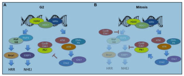
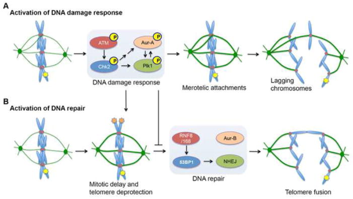
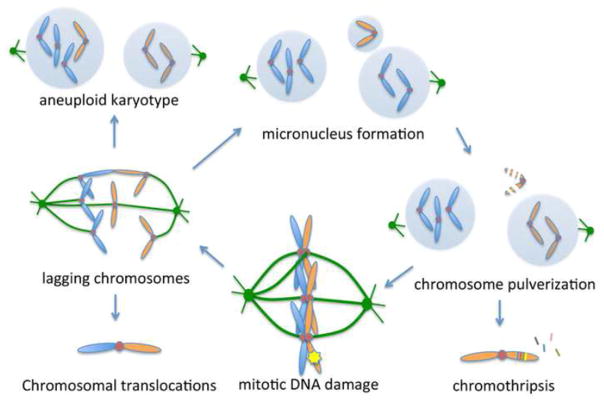
DNA double-strand breaks (DSBs) prevent cells from entering mitosis allowing cells to repair their genomic damage. Little is known about the response to DSBs once cells have already committed to mitosis. Here, we review the genome-protective role of the mitotic DNA damage response (DDR) and evidence suggesting that its untimely activation induces chromosome segregation errors and paradoxically undermines genomic integrity. In contrast to normal cells, cancer cells coopt this pathway to propagate structural and numerical chromosomal instabilities. Cells derived from genomically unstable tumors exhibit evidence for a partially activated DDR during mitosis, which leads to ongoing chromosome segregation errors. Thus, a thorough understanding of the consequences of mitotic DNA damage is key to our ability to devise novel anticancer therapeutic strategies.


Conflict of interest statement

Competing financial interests. The authors declare no competing financial interests.

Figures

Figure 1
Figure 1. Differences in the DNA damage response between mitosis and the G2 phase of the cell cycle
(A) Double strand DNA breaks (DSBs) elicit the phosphorylation of the modified histone H2AX (γ-H2AX) as well as the recruitment of mediator of the DNA damage checkpoint 1 (MDC1) and members of the MRN (Mre11-Rad50-Nbs1) complex. This, in turn leads to the activation of the transducers Ataxia-Telangiectasia mutated (ATM), ATR and DNA protein kinase (DNA-PK) and subsequent activation of Chk1 and Chk2 kinases. This leads to the inhibition of polo-like kinase 1 (Plk1) through the degradation of Bora and activation of the G2/M checkpoint preventing the onset of mitosis to allow time for DNA repair. Furthermore, RNF8 and RNF168 interact with MDC1, which allows their recruitment to DSB foci where they mediate the ubiquitination of γ-H2AX. This ubiquitination is key for the recruitment of Brca1 and 53BP1 to DSB foci and initiation of homologous recombination (HR)-mediated DNA repair and non-homologous end-joining (NHEJ), respectively. (B) During mitosis, induction of DSBs still leads to the immediate recruitment of γ-H2AX, MDC1, and members of the MRN complex. Mitotic cells also appear to exhibit activation of DNA damage transducers, however instead of inhibiting Plk1 evidence suggests that DNA damage response activation instead increases the levels of active phosphorylated Plk1. Importantly, mitotic cells inhibit DNA repair pathways though cyclin B/Cdk1-dependent, and mitosis specific, phosphorylation of Threonine-198 on RNF8, which prevents its interaction with MDC1 and recruitment to DSB foci. Furthermore, CDK1 and Plk1 phosphorylate 53BP1 on Threonine-1609 and Threonine-1618 preventing its binding to γ-H2AX. These two, mitosis specific phosphorylation events, effectively eliminate the recruitment of Brca1 and 53BP1 to DSBs and abrogate HR-mediated repair and NHEJ.
Figure 2
Figure 2. Consequences of activating the DNA damage response and DNA repair during mitosis
(A) Induction of DNA double-strand breaks (DSBs, depicted by yellow star on chromosome arm) during mitosis elicits a partial DNA damage response. The ATM/Chk2 arm of this mitotic DNA damage response was shown to increase kinetochore-microtubule (kMT) attachment stability by activating the mitotic kinases Aurora-A and Plk1. Increased kMT attachment stability increases the probability that erroneous kMT attachments (termed merotelic attachments) persist until anaphase onset. In this scenario, individual chromatids are simultaneously attached to microtubules emanating from opposite spindle poles and when these attachments persist until anaphase, they produce lagging chromosomes, which are a hallmark of whole-chromosome mis-segregation and numerical chromosomal instability. (B) In addition, the mitotic DNA damage response also leads to delay in mitotic progression, which was shown to lead to the deprotection of telomeres (depicted by orange stars on chromosome ends). Interestingly, however, the mitotic DNA damage response inhibits NHEJ and it was recently shown that ectopic activation of NHEJ leads to Aurora-B-mediated fusion of deprotected telomeres. Consequently, this can lead to dicentric chromosomes during anaphase and structural chromosomal instability.
Figure 3
Figure 3. The mitotic DNA damage response links structural and numerical cancer chromosome instabilities
By inducing chromosome segregation defects, mitotic DNA damage can lead to telomere fusions and structural aberrations leading to chromosome translocations. Lagging chromosomes can missegregate leading to aneuploidy or alternatively can be sequestered into micronuclei which are defective in DNA replication and lead to pulverization of their enclosed chromosome. This widespread structural chromosomal damage can persist into the subsequent mitosis leading to continued cycle of genomic instability. Alternatively, pulverized chromosomes can be repaired in a random fashion leading to focal complex rearrangements and deletions, a process known as chromothripsis.

References

    1. Gunderson LL, Tepper JE. Clinical Radiation Oncology. Churchill Livingstone; 2011.
    1. Sinclair WK. Cyclic x-ray responses in mammalian cells in vitro. Radiat Res. 1968;33:620–643. - PubMed
    1. Sinclair WK, Morton RA. X-ray sensitivity during the cell generation cycle of cultured Chinese hamster cells. Radiat Res. 1966;29:450–474. - PubMed
    1. Giunta S, et al. DNA damage signaling in response to double-strand breaks during mitosis. The Journal of Cell Biology. 2010;190:197–207. - PMC - PubMed
    1. Burrell RA, et al. Replication stress links structural and numerical cancer chromosomal instability. Nature. 2013;494:492–496. - PMC - PubMed

Publication types

LinkOut - more resources