

Enhancement of mTOR signaling contributes to acquired X-ray and C-ion resistance in mouse squamous carcinoma cell line
- PMID: 28718972
- PMCID: PMC5623753
- DOI: 10.1111/cas.13323
Enhancement of mTOR signaling contributes to acquired X-ray and C-ion resistance in mouse squamous carcinoma cell line
Abstract
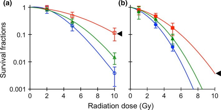
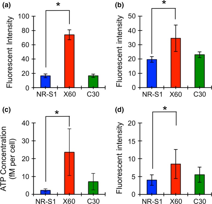
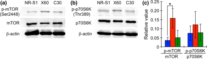
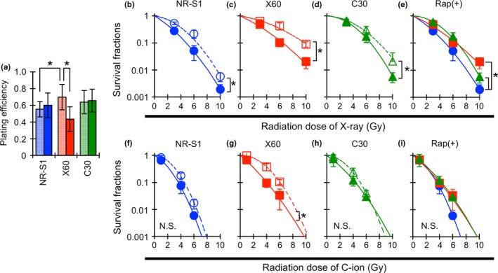
Our aim was to evaluate whether repetition of C-ion (carbon ion beam) irradiation induces radioresistance as well as repeated X-ray irradiation in cancer cell lines, and to find the key molecular pathway for radioresistance by comparing radioresistant cancer cells with their parental cells. A mouse squamous cell carcinoma cell line, NR-S1, and radioresistant cancer cells, NR-S1-C30 (C30) and NR-S1-X60 (X60), established by repetition of C-ion and X-ray irradiation, respectively, were used. X-ray and C-ion sensitivity, changes in lysosome, mitochondria, intracellular ATP and reactive oxygen species (ROS) level, and mechanistic target of rapamycin (mTOR) signaling were evaluated. Moreover, the effect of rapamycin on radioresistance was also assessed. X-ray and C-ion resistance of C30 cells was moderate, and the resistance of X60 cells was the highest in this study. In X60 cells, the amount of lysosome, mitochondria, intracellular ATP and ROS level were significantly increased, and mTOR and p70S6K (ribosomal protein S6 kinase p70) phosphorylation were enhanced compared with C30 and NR-S1 cells. The inhibition of mTOR signaling was effective for X-ray and C-ion radiosensitization in both cell lines, especially in X60 cells in which X-ray and C-ion resistance was decreased to the same level as that in NR-S1 cells. Our results indicated that the contribution to generate X-ray and C-ion resistance was less for repeated C-ion irradiations compared with repeated X-ray irradiation. Moreover, we found that activated mTOR signaling contributes to X-ray and C-ion resistance in the X60 cancer cells.
Keywords: Acquired radioresistance; energy metabolism; mTOR signaling; rapamycin; repeated X-ray and C-ion irradiations.
© 2017 The Authors. Cancer Science published by John Wiley & Sons Australia, Ltd on behalf of Japanese Cancer Association.
Figures
References
-
- Nagata Y, Hiraoka M, Shibata T et al Prospective trial of stereotactic body radiation therapy for both operable and inoperable T1N0M0 non‐small cell lung cancer: Japan Clinical Oncology Group Study JCOG0403. Int J Radiat Oncol Biol Phys 2015; 93: 989–96. - PubMed
-
- Miyamoto T, Baba M, Sugane T et al Carbon ion radiotherapy for stage I non‐small cell lung cancer using a regimen of four fractions during 1 week. J Thorac Oncol 2007; 2: 916–26. - PubMed
-
- Onishi H, Shirato H, Nagata Y et al Hypofractionated stereotactic radiotherapy (HypoFXSRT) for stage I non‐ small cell lung cancer: updated results of 257 patients in a Japanese multi‐ institutional study. J Thorac Oncol 2007; 2(Suppl 3): S94–100. - PubMed
-
- Koto M, Miyamoto T, Yamamoto N, Nishimura H, Yamada S, Tsujii H. Local control and recurrence of stage I non‐small cell lung cancer after carbon ion radiotherapy. Radiother Oncol 2004; 71: 147–56. - PubMed
-
- Kamada T, Tsujii H, Tsuji H et al Efficacy and safety of carbon ion radiotherapy in bone and soft tissue sarcomas. J Clin Oncol 2002; 20: 4466–71. - PubMed
Publication types
MeSH terms
Substances
LinkOut - more resources
Full Text Sources
Other Literature Sources
Molecular Biology Databases
Miscellaneous
