Review
. 2017 Sep;33(9):616-628.
doi: 10.1016/j.tig.2017.06.004. Epub 2017 Jul 15.

Left-Right Patterning: Breaking Symmetry to Asymmetric Morphogenesis

Affiliations
Review

Left-Right Patterning: Breaking Symmetry to Asymmetric Morphogenesis

Daniel T Grimes et al. Trends Genet. 2017 Sep.

Abstract

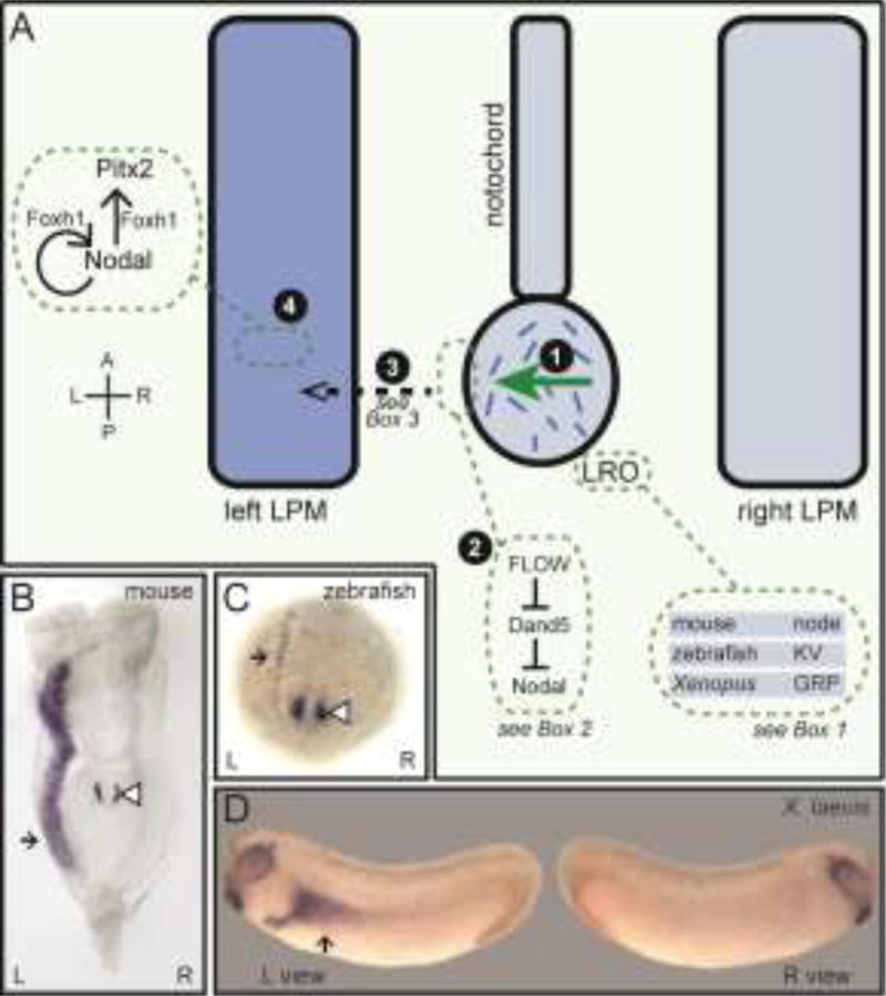
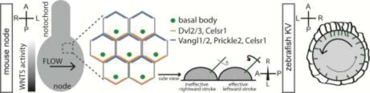
Vertebrates exhibit striking left-right (L-R) asymmetries in the structure and position of the internal organs. Symmetry is broken by motile cilia-generated asymmetric fluid flow, resulting in a signaling cascade - the Nodal-Pitx2 pathway - being robustly established within mesodermal tissue on the left side only. This pathway impinges upon various organ primordia to instruct their side-specific development. Recently, progress has been made in understanding both the breaking of embryonic L-R symmetry and how the Nodal-Pitx2 pathway controls lateralized cell differentiation, migration, and other aspects of cell behavior, as well as tissue-level mechanisms, that drive asymmetries in organ formation. Proper execution of asymmetric organogenesis is critical to health, making furthering our understanding of L-R development an important concern.

Keywords: Nodal; Pitx2; cell migration; cilia; left–right asymmetry; morphogenesis.


Figures

Figure 1
Figure 1. The origin of left-right asymmetry in vertebrates
(A) Embryonic L-R symmetry is broken during somite stages when an asymmetric fluid flow is generated by motile cilia within midline L-R coordinators (LROs), transient structures at the posterior end of the notochord (1). This asymmetric flow is sensed at the periphery of LROs, resulting in repression of Dand5, and thereby the activation of Nodal, on the left side (2). Asymmetric signals, possibly Nodal itself, are then propagated to LPM tissue (3) where Nodal signals are established in the left but not right LPM; there they activate further Nodal expression and expression of the transcription factor Pitx2, requiring FoxH1 (4). This asymmetric Nodal-Pitx2 pathway is critical for downstream asymmetric organ morphogenesis and is conserved across species. (B–D) Whole mount in situ hybridization in different organisms using probes for Nodal (B–C) or Pitx2 (D). Black arrow denotes LPM expression while white arrowhead denotes LRO expression. A; anterior, GRP; gastroceol roof plate, KV; Kupffer's vesicle, L; left, LPM; lateral plate mesoderm, LRO; left-right coordinator, R; right, P; posterior.
Key Figure. Figure 2
Key Figure. Figure 2. L-R asymmetric organogenesis of the gut, heart, and brain
(A) Upper panels: the endoderm (green) acquires an asymmetric morphology between 24 and 30 hpf. By 30 hpf, the liver (L) and pancreatic (P) buds have emerged and the intestine has looped asymmetrically. Lower panels: at 24 hpf, the gut endoderm exists as a central rod surrounded by LPM. The LPM (orange) migrates medially toward the endodermal rod, with the left LPM migrating dorsally and the right LPM ventro-laterally. By 30 hpf, the gut has shifted to the left as a result of these LPM migrations. (B) In amniotes, the gut tube is connected to the body wall by the dorsal mesentery. Cell shape, adhesion, and ECM changes downstream of the Nodal-Pitx2 pathway, with the right mesenchyme decondensing and the epithelium attaining a cuboidal architecture, result in the tilting of the gut tube towards the left, a critical first asymmetry in the gut looping process. Boxed panel: differential elongation of the DM and the gut tube cause compressive forces to be imparted on the gut (purple arrows), resulting in a buckling event that drives looping. (C) Schematic of mouse lungs showing four right-sided lobes and a single left-sided lung lobe. Bilateral Nodal-Pitx2 pathway activity in mutants generally leads to left lung isomerism whereas right isomerism occurs in mutants that fail to activate Nodal-Pitx2. Examples of mutants displaying these phenotypes are given. (D) Heart jogging in zebrafish occurs when a symmetrical cardiac cone, composed of atrial (light red) and ventricular (dark red) cells, a volcano-shaped structure (see side view in dotted box), becomes asymmetric to form the cardiac tube by 24 hpf. Spaw signals from the left LPM, which activates target genes in the left-sided cells of the cone only. Subsequently, the left cells migrate left and anterior at a faster rate than cells on the right. This results in the clockwise rotation and, owing to involution and extension extension of the cone, forms a linear tube that points to the left side. (E) Schematic of the epithalamic region of the zebrafish brain showing the left and right Hb, the PpO and the ep. The PpO migrates left towards the left Hb down an FGF gradient that increases from the midline to the lateral sides. Nodal, active on the left side only, promotes left-sided migration of the PpO. A; anterior, D; dorsal, ep; epiphysis, Hb; habenulae, IL; inferior lobe, L; left, LL; left lobe, LPM; lateral plate mesoderm, ML; middle lobe, P; posterior, PCL; post-caval lobe, PpO; parapineal organ, R; right, SL; superior lobe, V; ventral.
None
None

Similar articles

Cited by

References

    1. Shiraishi I, Ichikawa H. Human heterotaxy syndrome - from molecular genetics to clinical features, management, and prognosis. Circulation journal : official journal of the Japanese Circulation Society. 2012;76:2066–2075. - PubMed
    1. Kennedy MP, Omran H, Leigh MW, Dell S, Morgan L, Molina PL, Robinson BV, Minnix SL, Olbrich H, Severin T, et al. Congenital heart disease and other heterotaxic defects in a large cohort of patients with primary ciliary dyskinesia. Circulation. 2007;115:2814–2821. - PubMed
    1. Hachisuga M, Oki S, Kitajima K, Ikuta S, Sumi T, Kato K, Wake N, Meno C. Hyperglycemia impairs left-right axis formation and thereby disturbs heart morphogenesis in mouse embryos. Proceedings of the National Academy of Sciences of the United States of America. 2015;112:E5300–5307. - PMC - PubMed
    1. Shiratori H, Hamada H. TGFbeta signaling in establishing left-right asymmetry. Seminars in cell & developmental biology. 2014;32:80–84. - PubMed
    1. Amack JD. Salient features of the ciliated organ of asymmetry. Bioarchitecture. 2014;4:6–15. - PMC - PubMed

Publication types