. 2017 Sep 15;28(19):2543-2554.
doi: 10.1091/mbc.E17-01-0068. Epub 2017 Jul 18.

Dynein efficiently navigates the dendritic cytoskeleton to drive the retrograde trafficking of BDNF/TrkB signaling endosomes

Affiliations

Dynein efficiently navigates the dendritic cytoskeleton to drive the retrograde trafficking of BDNF/TrkB signaling endosomes

Swathi Ayloo et al. Mol Biol Cell. .

Abstract

The efficient transport of cargoes within axons and dendrites is critical for neuronal function. Although we have a basic understanding of axonal transport, much less is known about transport in dendrites. We used an optogenetic approach to recruit motor proteins to cargo in real time within axons or dendrites in hippocampal neurons. Kinesin-1, a robust axonal motor, moves cargo less efficiently in dendrites. In contrast, cytoplasmic dynein efficiently navigates both axons and dendrites; in both compartments, dynamic microtubule plus ends enhance dynein-dependent transport. To test the predictions of the optogenetic assay, we examined the contribution of dynein to the motility of an endogenous dendritic cargo and found that dynein inhibition eliminates the retrograde bias of BDNF/TrkB trafficking. However, inhibition of microtubule dynamics has no effect on BDNF/TrkB motility, suggesting that dendritic kinesin motors may cooperate with dynein to drive the transport of signaling endosomes into the soma. Collectively our data highlight compartment-specific differences in kinesin activity that likely reflect specialized tuning for localized cytoskeletal determinants, whereas dynein activity is less compartment specific but is more responsive to changes in microtubule dynamics.


Figures

FIGURE 1:
FIGURE 1:
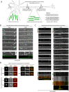
Recruitment of dynein or kinesin to peroxisomes in axons and dendrites. (A) Schematic and parts list of the light-inducible dimerization system implemented in mature neurons that have uniformly polarized microtubule arrays in the axon and mixed arrays in dendrites. (B) Time series and corresponding kymographs showing the anterograde movement of locally photoactivated peroxisomes (white box) in axons. Horizontal bar, 5 µm. Vertical bar, 1 min. (C) Time series and corresponding kymographs showing the bidirectional movement of locally photoactivated peroxisomes (white box) in dendrites. Horizontal bar, 5 µm. Vertical bar, 1 min. (D) Top panel, images of motor recruitment to peroxisomes pre- and postphotoactivation. Horizontal bar, 1 µm. Bottom panel, representative stills showing recruitment of K560 even in the case of peroxisomes that are immotile postphotoactivation. Horizontal bar, 500 nm.
FIGURE 2:
FIGURE 2:
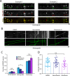
Kinesin-1 moves inefficiently while dynein has a retrograde bias in dendrites. (A) Percentage of photoactivated peroxisomes that are motile in dendrites. Mean ± SEM, **p < 0.01, ***p < 0.001, Student’s t test. (B) Average velocity of motile photoactivated peroxisomes following dynein or K560 recruitment. Mean ± SEM, n.s. = not significant, Student’s t test. (C–F) Histograms of run length and run time of the individual runs of motile peroxisomes following recruitment of dynein (C, D) or K560 (E, F). (G) Quantitation of the overall percent of peroxisome runs in the anterograde vs. retrograde direction upon motor recruitment. Data from 25 peroxisomes from n = 12 neurons and N = 3 independent experiments. (H) Quantitation of directionality of EB3 comets in dendrites and representative kymographs in the axon and dendrites of mature hippocampal neurons, 8–10 DIV. Mean ± SEM, n = 10 neurons from N = 2 independent experiments. Horizontal bar, 5 µm. Vertical bar, 30 s. (I) Quantitation of percent of peroxisomes going back to the cell body postphotoactivation. Mean ± SEM, **p < 0.01, Student’s t test. Data from 25 peroxisomes from n = 12 neurons and N = 3 independent experiments. (J) Representative examples showing bidirectional movement of peroxisomes upon K560 recruitment or going back to the cell body upon dynein recruitment. Horizontal bar, 5 µm. Vertical bar, 1 min.
FIGURE 3:
FIGURE 3:
Low-dose nocodazole treatment decreases tubulin tyrosination. (A) Representative deconvolved maximum projections of STED images of tyrosinated tubulin in hippocampal neurons with and without low-dose nocodazole treatment. (B) Axonal and (C) dendritic quantification of tyrosinated tubulin. Tyrosinated tubulin levels were normalized to β-tubulin levels and represented relative to the DMSO group. Mean ± SEM. ***p < 0.001, Mann-Whitney U test; n = 22–37 neurons in each group from N = 2 independent experiments. (D) Representative deconvolved maximum projections of STED images of detyrosinated tubulin in hippocampal neurons with and without nocodazole treatment. (E) Axonal and (F) dendritic quantification of detyrosinated tubulin. Detyrosinated tubulin levels were normalized to β-tubulin levels and represented relative to the DMSO group. **p < 0.01, ***p < 0.001, Mann-Whitney U test; n = 28–29 neurons from N = 2 independent experiments. (G) Axonal and dendritic quantification of β-tubulin in neurons immunostained for tyrosinated α-tubulin, used for quantification B and C. β-Tubulin levels are represented relative to β-tubulin levels in the DMSO group. n.s., not significant (p > 0.05), Mann-Whitney U test; n = 22–37 neurons in each group from N = 2 independent experiments. (H) Axonal and dendritic quantification of β-tubulin in neurons immunostained for detyrosinated α-tubulin, used for quantification in E and F. β-Tubulin levels are represented relative to β-tubulin levels in the DMSO group. n.s., not significant (p > 0.05), Mann-Whitney U test; n = 28–29 neurons in each group from N = 2 independent experiments.
FIGURE 4:
FIGURE 4:
Dynein requires dynamic microtubules for efficient transport in axons and dendrites. (A) Representative kymographs of EB3 comets in axons and dendrites of hippocampal neurons treated with DMSO or 100 ng/ml nocodazole for 1.5 h at 37°C. Horizontal bar, 5 µm. Vertical bar, 30 s. (B) Representative kymographs showing movement of photoactivated peroxisomes in axons. Horizontal bar, 5 µm. Vertical bar, 1 min. (C) Quantitation of percentage of peroxisomes that are motile in axons of neurons expressing K560, treated with DMSO or nocodazole. Mean ± SEM. (D) Quantitation of percentage of peroxisomes that are motile in axons of neurons expressing BICD, mean ± SEM, (E) their average velocities, mean ± SD, and (F) number of pauses per photoactivated peroxisome, mean ± SEM. (G, H) Quantitation of percentage of peroxisomes that are motile in dendrites of neurons expressing K560 or BICD. Mean ± SEM. (I) With reduced transport in dendrites, there is a concomitant decrease in percentage of peroxisomes going back to the cell body in the case of BICD. Data from 15–20 peroxisomes, n = 14 neurons for axons and 25–30 peroxisomes, n = 16 neurons for dendrites from N = 3 independent experiments, n.s., not significant, *p < 0.05, **p < 0.01, Student’s t test in E and H, one-way ANOVA with Tukey’s post hoc test in the rest.
FIGURE 5:
FIGURE 5:
Dynein contributes to the retrograde transport of TrkB in dendrites. (A) Representative images of TrkB puncta in cell soma and dendrites of hippocampal neurons. Horizontal bar, 5 µm. (B) Representative kymographs showing the movement of TrkB-mRFP in neurons treated with DMSO or ciliobrevin D or nocodazole. (C) Fraction of TrkB puncta exhibiting anterograde or retrograde movement. Mean ± SEM. (D) Average velocities of motile TrkB puncta. Mean ± SD. Data from n = 20 neurons from N = 3 independent experiments. *p < 0.05, n.s., not significant, Student’s t test.
FIGURE 6:
FIGURE 6:
BDNF/TrkB motility in dendrites is altered by dynein inhibition. (A) Colocalization of TrkB-RFP with BDNF-Qdots in hippocampal neuron dendrites. Horizontal bar, 5 µm. (B) Representative kymographs showing the movement of BDNF-Qdots in neurons treated with DMSO or ciliobrevin D or nocodazole. (C) Fraction of BDNF-Qdots exhibiting anterograde or retrograde movement. Mean ± SEM. *p < 0.05, n.s., not significant, one-way ANOVA with Tukey’s post hoc test. (D) Average velocities of motile BDNF-Qdots. Mean ± SD. Data from n = 15–18 neurons from N = 3 independent experiments; n.s., not significant, Student’s t test.
FIGURE 7:
FIGURE 7:
Working model for the axo-dendritic regulation of motor proteins. Microtubules are differentially organized in the axon and dendrites of mammalian neurons (shown in shades of green). Dynein (purple) motors recruited to peroxisomes using photoactivation demonstrate a retrograde bias within dendrites that reflects the overall organization of the microtubule cytoskeleton in this compartment, as 60% of microtubules are oriented with minus ends toward the soma in mammalian dendrites. In contrast, kinesin-1 (blue) motors recruited to peroxisomes show no overall directional bias in dendrites. Furthermore, efficient dynein motility requires dynamic, tyrosinated microtubules (red), whereas kinesin motility is unaffected by microtubule dynamics.

References

    1. Arpag G, Shastry S, Hancock WO, Tuzel E. Transport by populations of fast and slow kinesins uncovers novel family-dependent motor characteristics important for in vivo function. Biophys J. 2014;107:1896–1904. - PMC - PubMed
    1. Ayloo S, Lazarus JE, Dodda A, Tokito M, Ostap EM, Holzbaur EL. Dynactin functions as both a dynamic tether and brake during dynein-driven motility. Nat Commun. 2014;5:4807. - PMC - PubMed
    1. Baas PW, Deitch JS, Black MM, Banker GA. Polarity orientation of microtubules in hippocampal neurons: uniformity in the axon and nonuniformity in the dendrite. Proc Natl Acad Sci USA. 1988;85:8335–8339. - PMC - PubMed
    1. Ballister ER, Aonbangkhen C, Mayo AM, Lampson MA, Chenoweth DM. Localized light-induced protein dimerization in living cells using a photocaged dimerizer. Nat Commun. 2014;5:5475. - PMC - PubMed
    1. Ballister ER, Ayloo S, Chenoweth DM, Lampson MA, Holzbaur EL. Optogenetic control of organelle transport using a photocaged chemical inducer of dimerization. Curr Biol. 2015;25:R407–R408. - PMC - PubMed

MeSH terms