. 2017 Aug 1;114(31):8295-8300.
doi: 10.1073/pnas.1703001114. Epub 2017 Jul 18.

Broadly expressed repressors integrate patterning across orthogonal axes in embryos

Affiliations

Broadly expressed repressors integrate patterning across orthogonal axes in embryos

Theodora Koromila et al. Proc Natl Acad Sci U S A. .

Abstract

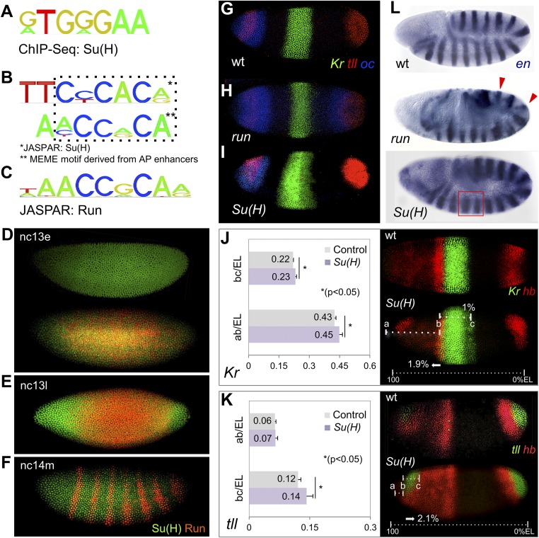
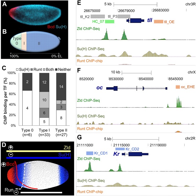
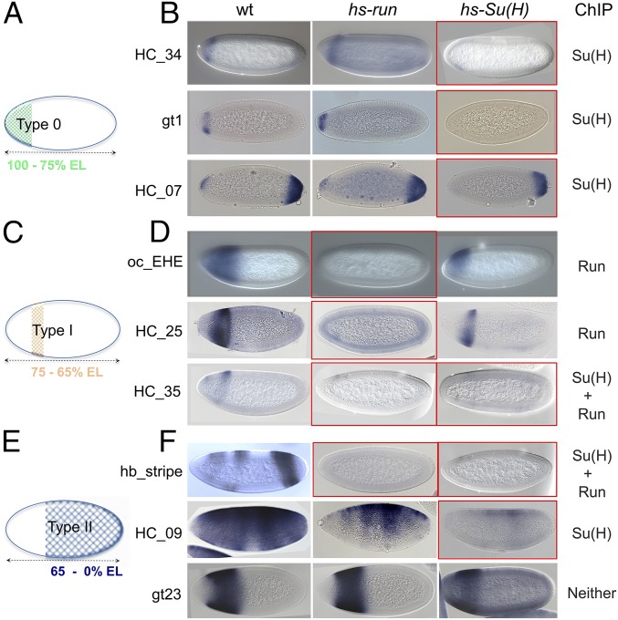
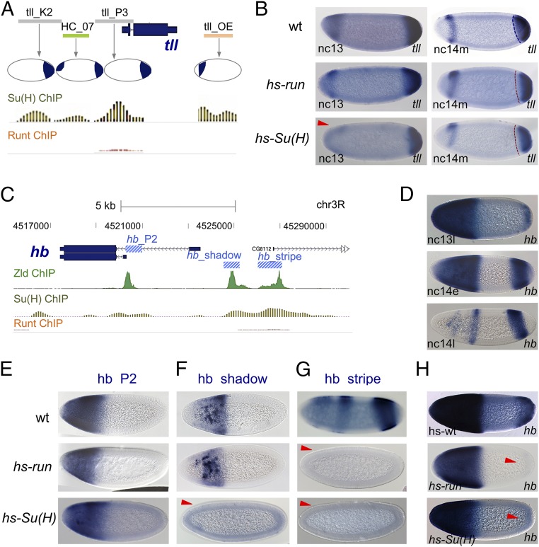
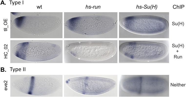
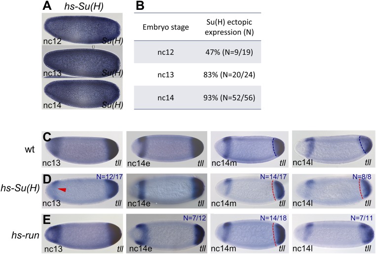
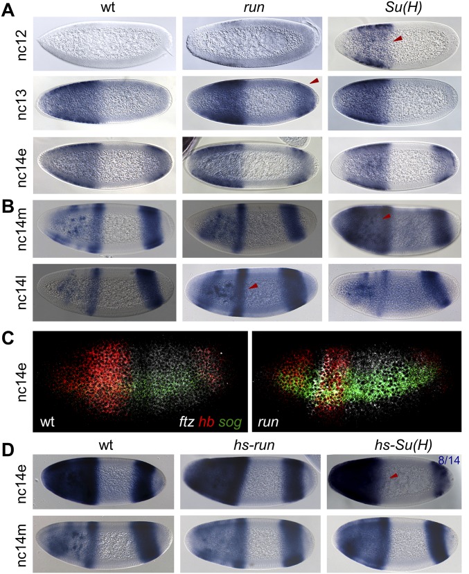
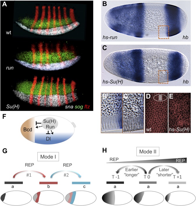
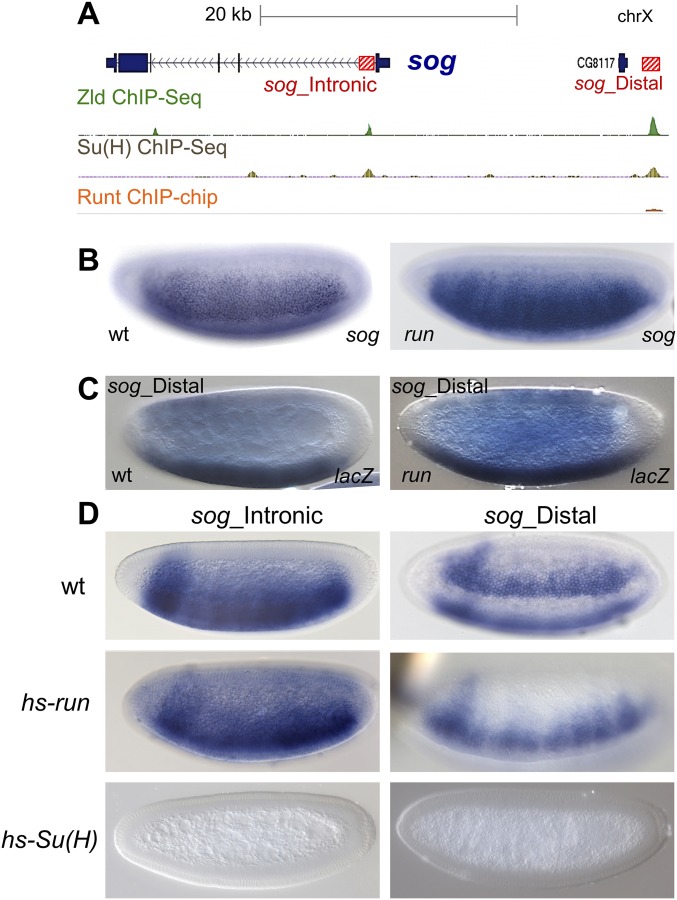
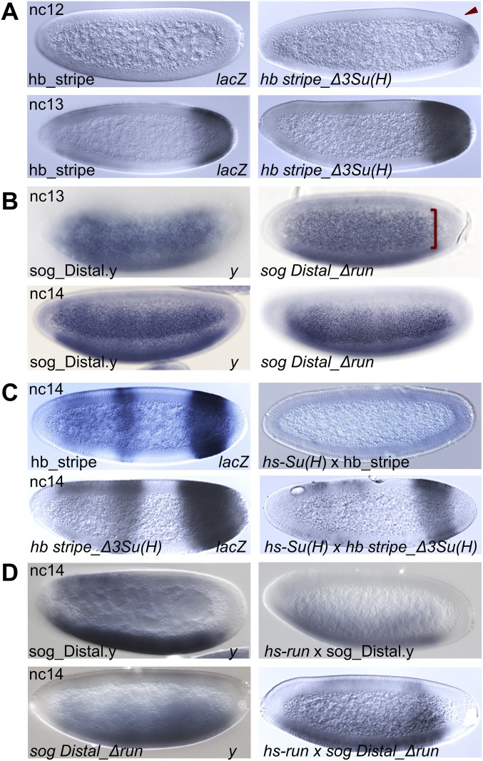
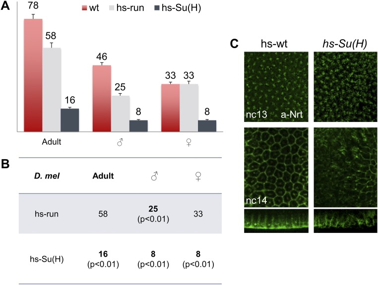
The role of spatially localized repressors in supporting embryonic patterning is well appreciated, but, alternatively, the role ubiquitously expressed repressors play in this process is not well understood. We investigated the function of two broadly expressed repressors, Runt (Run) and Suppressor of Hairless [Su(H)], in patterning the Drosophila embryo. Previous studies have shown that Run and Su(H) regulate gene expression along anterior-posterior (AP) or dorsal-ventral (DV) axes, respectively, by spatially limiting activator action, but here we characterize a different role. Our data show that broadly expressed repressors silence particular enhancers within cis-regulatory systems, blocking their expression throughout the embryo fully but transiently, and, in this manner, regulate spatiotemporal outputs along both axes. Our results suggest that Run and Su(H) regulate the temporal action of enhancers and are not dedicated regulators of one axis but, instead, act coordinately to pattern both axes, AP and DV.

Keywords: Runt; Su(H); embryonic patterning; enhancers; transcriptional repressor.


Conflict of interest statement

The authors declare no conflict of interest.

Figures

Fig. 1.
Fig. 1.
Su(H) binding site and mutant phenotypes suggest a role for Su(H) in AP patterning. In this and all subsequent figures, Drosophila embryo images are depicted with anterior to the left and dorsal up unless otherwise noted. (AC) Comparison of Su(H) DNA binding consensus site derived from ChIP-seq (A; 24% of called peaks compared with 6% background) (7), Drosophila Su(H) motif from JASPAR (B, Top; reverse complement relative to A), motif overrepresented within AP enhancers (B, Bottom) (6), and Drosophila Run consensus binding site (C) (6). (DF) Anti-Su(H) (green) and anti-Run (red) protein staining of embryos at early stage nc 13 (nc13e; D), late nc 13 (nc13l; E), and mid-nc 14 (nc14m; F). (GI) FISH using riboprobes to detect Kr (green), tll (red), and oc (blue) transcripts in WT embryos (G) as well as in run (H) and Su(H) (I) mutant embryos. (J and K) Ratio of Kr and tll transcript expression domains relative to total EL. Embryos processed by FISH using Kr, hb, and/or tll riboprobes to detect transcripts in WT and Su(H) mutants at early nc 14. The anterior boundaries of the central Kr (J) or anterior tll (K) domains are marked as “ab,” whereas “bc” demarcates the central, dorsal, Kr domain width (J), or anterior tll domain length (K). (L) en transcript expression in germband-elongated embryos of WT, run mutant, or Su(H) mutant genetic backgrounds. Red arrowheads mark odd en stripes in run embryos, whereas a red box indicates the en interstripe distance in Su(H) mutants.
Fig. S1.
Fig. S1.
Ectopic expression of Runt and Su(H) impacts En expression in germband-elongated embryos. Germband-elongated embryos are oriented with anterior aspect to the left. In situ hybridization using a riboprobe to en shows expression within 14 stripes in WT embryos undergoing germband elongation (A). Upon ectopic expression of Runt, en is down-regulated in odd-numbered stripes (B; even stripes marked by arrowhead), whereas ectopic expression of Su(H) has a distinct phenotype resulting in an increase in interstripe distance (C, red box). hs-Su(H) and Su(H) mutant embryos exhibit opposite phenotypes: compare red boxes in C and in Fig. 1L.
Fig. 2.
Fig. 2.
Transcription factors binding to three different enhancer types that drive expression along the AP axis. (A) Su(H) (cyan) and Bicoid (Bcd; red) protein expression in early nc 14 embryos. (B) Schematics of three Bcd-defined enhancer types, type 0 (green), type I (orange), and type II (light blue), classified by the positions of their Bcd-dependent posterior boundaries based on Chen et al. (6). Left end of schematic represents anterior tip of the embryo (100% EL). (C) A total of 66 Bcd-dependent enhancers derived from Chen et al. (6) were classified based on whether endogenous sequences exhibit Su(H) and/or Run ChIP binding or a lack thereof (“Neither”). (D) Representation of Zld, Su(H), Bcd, and Run expression patterns within a schematic of an early embryo. Colored lines represent regions of expression for each of these factors. “+” defines input by activators Zld and Bcd, and “−“ defines input by repressors Su(H) and Run. (EG) ChIP binding data for Zelda (13), Su(H) (7), and Run (14) showing occupancy of these factors at three loci: tll (E), oc (F), and Kr (G). Location for previous characterized enhancers active in the early embryo are shown and colored if associated with type 0 (green), type I (orange), or type II (blue) AP enhancers (B) or none (gray).
Fig. 3.
Fig. 3.
Ectopic expression of Su(H) or Runt has variable effects on AP enhancers. (A, C, and E) Schematic of domains of expression for gap genes expressed along the AP axis of Drosophila embryos that fall into the type 0 (A), type I (C), or type II (E) enhancer categories. (B, D, and F) Embryos containing the indicated enhancer constructs of type 0 (B), type I (D), or type II (F) classification within three different genetic backgrounds, WT embryos, hs-run, and hs-Su(H), were equivalently heat-shocked, and lacZ reporter expression examined by in situ hybridization. Embryos outlined in red from experiments showing loss of expression associated with heat shock. ChIP-defined binding to these enhancers sequences (endogenous locations) as assayed for occupancy by Su(H), Run, Su(H) + Run, or neither factor is shown (Right).
Fig. 4.
Fig. 4.
Broadly expressed repressors affect gap gene patterns via impacting the timing of action for a subset of their enhancers. (A) Relative location of four tll enhancer sequences (tll_K2, HC_07, tll_P3, tll_OE) to tll gene as well as the pattern of expression supported by each enhancer diagramed within the embryo schematics (26) compared with Su(H) and Run ChIP-defined in vivo occupancy to these sequences. (B) tll gene expression in heat-shocked WT and hs-run or hs-Su(H) embryos at nc 13 and mid-nc 14. (C) ChIP data for Zelda, Su(H), and Run binding to the hb locus relative to position of three enhancers (blue boxes) (19), supporting early embryonic hb expression. (D) Endogenous hb expression at three stages in the early embryo detected by in situ hybridization shows that the pattern is very dynamic. (EH) Expression associated with hb enhancer reporter constructs using lacZ riboprobe (EG) or endogenous hb (H) in heat-shocked WT, hs-run, and hs-Su(H) embryos at mid-nc 14 (EG) or at the end of nc 13 (H). Delayed hb phenotype exhibited in 12 of 15 hs-run and 11 of 15 hs-Su(H) embryos (H). Red arrowheads mark domains where patterns exhibit alterations.
Fig. S2.
Fig. S2.
Runt and Su(H) ChIP occupancy of genomic enhancer sequences does not always correlate with the ability of these factors to repress reporter constructs. Expression patterns of some reporter genes from type I (A) and type II (B) enhancers (Left) in embryos collected from three different genetic backgrounds: WT, hs-run, or hs-Su(H). All embryos were equivalently processed, including heat shock of WT, which serves as control. Runt and Su(H) ectopic expression does not completely silence the expression of tll_OE, HC_02 (slp2 enhancer), and eve2 reporters, despite detection of ChIP binding to these sequences at their endogenous locations. No binding was detected to the eve2 locus.
Fig. S3.
Fig. S3.
tll expression at the anterior and posterior poles is dynamic and differentially impacted by hs-Su(H) and hs-run. (A) Hs-Su(H) embryos overexpressing Su(H) gene at different stages of early development. (B) Quantification of ectopic expression assay efficiency at different ncs (nc 12–14). (CE) Developmental time-course of tll expression within early embryos of indicated stages detected by in situ hybridization using tll riboprobes. All embryos were heat-shocked, including WT for comparative purposes. tll expression is shown for embryos of WT (C), hs-Su(H) (D), or hs-run (E) background at nc 13 until late nc 14. tll expression at the anterior and posterior poles is dynamic. Quantification of embryos that exhibit similar to the depicted phenotypes are present on the right corner of each image. Fifteen embryos were examined and the ratio was 14/15 embryos or greater if it is not presented. Red arrowhead and dashed lines indicate domains of reduced expression. SI Materials and Methods includes details about how tll posterior expression measurements were made.
Fig. S4.
Fig. S4.
Su(H) and Runt affect hb spatiotemporal patterning. (A and B) hb expression shown in WT, run, and Su(H) embryos detected by using in situ hybridization with an intronic hb riboprobe to embryos of nc 12, nc 13, and early nc 14 as indicated (A). Two more stages (mid-late nc 14) identified by using hb exon riboprobe (B). (C) FISH using riboprobes to ftz (gray), hb (red), and sog (green) to examine transcript expression domain in WT vs. run mutant embryos (late nc 13). (D) Endogenous hb expression in heat-shocked WT and hs-run or hs-Su(H) embryos at mid- and late nc 14. Red arrowhead indicates reduced expression upon ectopic expression of hs-SuH. Quantification of the depicted phenotypes are present in the right corner of each image unless the ratio is 14/15 embryos or greater.
Fig. 5.
Fig. 5.
Ubiquitous repressors regulate enhancer action across embryonic axes. (A) FISH using riboprobes to sna (white), sog (green), and ftz (red) show transcript expression domains within WT as well as run and Su(H) mutant embryos (mid-nc 14). (B and C) Ectopic expression of Su(H) through heat shock of hs-SuH embryos results in cellularization defects at late cycle 14 (C and C’). In contrast, no such cellularization phenotypes are observed upon heat shock of hs-run (B and B′) or WT embryos (Fig. S7C). (B′ and C′) Magnified views of B and C, respectively. (D and E) Fluorescent staining of embryos shows anomalous distribution of cell membranes within hs-Su(H) embryos (E) compared with WT (D) at mid-nc 14. Embryos in BE processed by in situ hybridization using hb (B and C) or Kr (D and E) probes. Although expression of these genes is not necessarily relevant to cellularization defects, this confirms that the embryos are fertilized and development had progressed. (F) Broadly acting transcription factors Su(H) and Run encompass multiple roles in patterning by acting as repressors to regulate gene expression along AP and DV axes together with Bicoid (Bcd) and/or Dorsal (Dl) morphogens, respectively. As these factors are known to exhibit dual function, their roles as activators may also be more widespread. (G and H) Two different mechanisms by which broadly expressed repressors may impact spatiotemporal patterning are depicted. Repressors may regulate the timing of action for different enhancers acting in series (G) or, alternatively, repressors may influence the length of time a single enhancer is active (H) to impact spatiotemporal outputs.
Fig. S5.
Fig. S5.
Assays to test the role for Runt, in addition to Su(H), in the regulation of the sog gene. (A) ChIP data for Zld, Su(H), and Runt showing occupancy at the sog gene locus. Position of two enhancers controlling sog early embryonic expression, sog_Intronic and sog_Distal, are indicated by red boxes. All three factors exhibit binding to sog_Distal, but only two of the three factors [Zld and Su(H)] exhibit binding to sog_Intronic. (BD) Expression of transcripts in embryos was obtained using in situ hybridization and specific riboprobes. (B) Expression of sog in WT or run mutant embryos at late nc 14. sog is expanded in run mutants. (C) Expression of lacZ in embryos, either WT or run mutants, containing the sog_Distal reporter at late nc 13. The reporter is more broadly expressed in the run mutant compared with WT embryos. (D) Expression of sog_Intronic or sog_Distal in WT, hs-run, and hs-Su(H) embryos that have been heat-shocked. Ectopic Su(H) represses expression of both reporters, suggesting a role for Su(H) in regulation of both sog enhancers. Alternatively, ectopic Runt was not able to repress expression of either reporter.
Fig. S6.
Fig. S6.
Mutagenesis of Su(H) and Run binding sites within enhancers demonstrates a direct role for these factors in the regulation of hb and sog gene expression. (A and B) Reporter gene expression in embryos of indicated stages driven by hb_stripe (A) and sog_Distal.y (B) WT or mutated enhancers assayed using riboprobes to reporters lacZ (A) or yellow (B). Three Su(H) sites (matches to the binding site consensus) were mutated in hb_stripe enhancer sequence [hb stripe_Δ3Su(H)], and one Run site was mutated in sog_Distal enhancer sequence [sogDistal_Δrun]. Mutation of Su(H) sites results in the h_stripe enhancer reporter supporting earlier expression relative to WT, whereas mutation of the Run site in the sog_Distal enhancer reporter results in expansion of expression at nc 13 but has little effect at nc 14. Red arrowhead and bracket indicate domains where patterns exhibit alterations. (C and D) Heat shock-mediated ectopic expression of Su(H) or Run on WT or mutated hb_stripe and sog_Distal enhancer sequences. Whereas ectopic expression of Su(H) is able to silence expression of the hb_stripe reporter, the hb stripe_Δ3Su(H) reporter was refractory to repression (C). Similarly, whereas ectopic expression of Run down-regulates expression of the sog_Distal.y reporter, the sogDistal_Δrun reporter was refractory to repression (D). The control enhancers [hb_stripe, hb stripe_Δ3Su(H), sog_Distal.y, sogDistal_Δrun] were similarly treated as the hs-Su(H) samples (i.e., heat-shocked).
Fig. S7.
Fig. S7.
Ectopic expression of Run or Su(H) within embryos results in decreased viability of adult flies, including sex-specific differences, as well as cellularization defects in embryos. (A) Flies survived after heat-shocking WT embryos, as well as embryos carrying hs-run or hs-Su(H) reporter constructs. The overall survival rate decreased to 20.5% in hs-Su(H) embryos. These data support a role for Runt in sex determination, as there was a 46% decrease in male progeny in the hs-run experiment. (B) P value analysis presented in B. (C) Anti-Nrt staining of early hs-WT and hs-Su(H) Drosophila embryos (nc 13 and nc 14) as indicated. Lateral views and cross-sections of nc 14 hs-Su(H) embryos showed cellularization defects compared with hs-WT of the same stage.

References

    1. Briscoe J, Small S. Morphogen rules: Design principles of gradient-mediated embryo patterning. Development. 2015;142:3996–4009. - PMC - PubMed
    1. Rushlow CA, Shvartsman SY. Temporal dynamics, spatial range, and transcriptional interpretation of the Dorsal morphogen gradient. Curr Opin Genet Dev. 2012;22:542–546. - PubMed
    1. Reeves GT, Stathopoulos A. Graded dorsal and differential gene regulation in the Drosophila embryo. Cold Spring Harbor Perspect Biol. 2009;1(4):a000836. - PMC - PubMed
    1. Sun Y, et al. Zelda overcomes the high intrinsic nucleosome barrier at enhancers during Drosophila zygotic genome activation. Genome Res. 2015;25:1703–1714. - PMC - PubMed
    1. Morel V, Schweisguth F. Repression by suppressor of hairless and activation by Notch are required to define a single row of single-minded expressing cells in the Drosophila embryo. Genes Dev. 2000;14:377–388. - PMC - PubMed

LinkOut - more resources