

Optical Coherence Tomography Findings in Patients With Coronary Stent Thrombosis: A Report of the PRESTIGE Consortium (Prevention of Late Stent Thrombosis by an Interdisciplinary Global European Effort)
- PMID: 28720725
- PMCID: PMC5598909
- DOI: 10.1161/CIRCULATIONAHA.117.026788
Optical Coherence Tomography Findings in Patients With Coronary Stent Thrombosis: A Report of the PRESTIGE Consortium (Prevention of Late Stent Thrombosis by an Interdisciplinary Global European Effort)
Abstract
Background: Stent thrombosis (ST) is a serious complication following coronary stenting. Intravascular optical coherence tomography (OCT) may provide insights into mechanistic processes leading to ST. We performed a prospective, multicenter study to evaluate OCT findings in patients with ST.
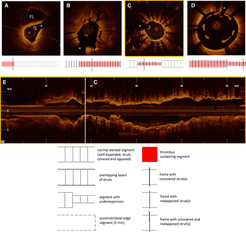
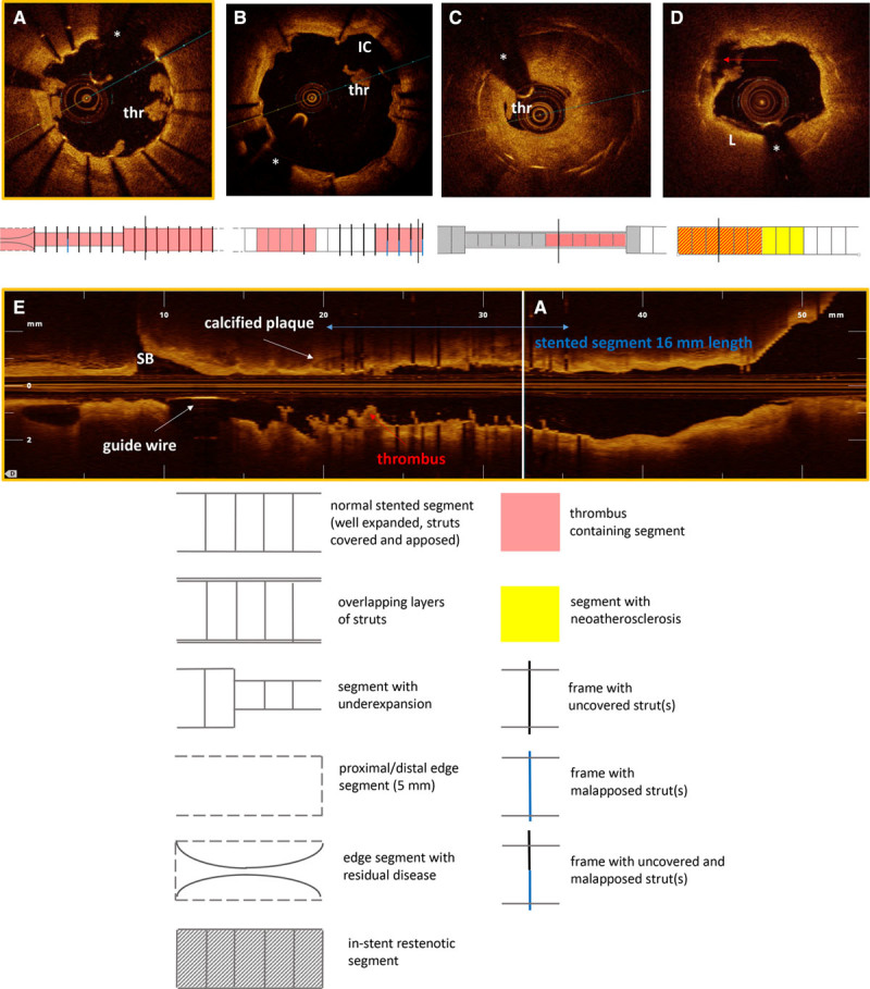
Methods: Consecutive patients presenting with ST were prospectively enrolled in a registry by using a centralized telephone registration system. After angiographic confirmation of ST, OCT imaging of the culprit vessel was performed with frequency domain OCT. Clinical data were collected according to a standardized protocol. OCT acquisitions were analyzed at a core laboratory. Dominant and contributing findings were adjudicated by an imaging adjudication committee.
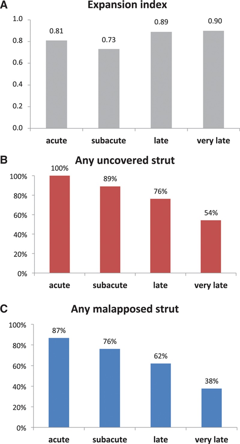
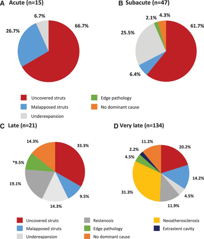
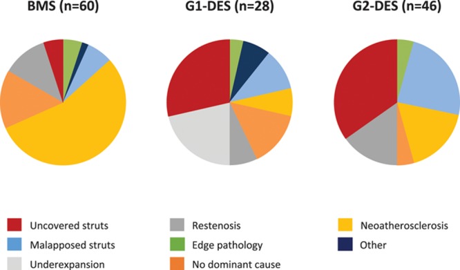
Results: Two hundred thirty-one patients presenting with ST underwent OCT imaging; 14 (6.1%) had image quality precluding further analysis. Of the remaining patients, 62 (28.6%) and 155 (71.4%) presented with early and late/very late ST, respectively. The underlying stent type was a new-generation drug-eluting stent in 50.3%. Mean reference vessel diameter was 2.9±0.6 mm and mean reference vessel area was 6.8±2.6 mm2. Stent underexpansion (stent expansion index <0.8) was observed in 44.4% of patients. The predicted average probability (95% confidence interval) that any frame had uncovered (or thrombus-covered) struts was 99.3% (96.1-99.9), 96.6% (92.4-98.5), 34.3% (15.0-60.7), and 9.6% (6.2-14.5) and malapposed struts was 21.8% (8.4-45.6), 8.5% (4.6-15.3), 6.7% (2.5-16.3), and 2.0% (1.2-3.3) for acute, subacute, late, and very late ST, respectively. The most common dominant finding adjudicated for acute ST was uncovered struts (66.7% of cases); for subacute ST, the most common dominant finding was uncovered struts (61.7%) and underexpansion (25.5%); for late ST, the most common dominant finding was uncovered struts (33.3%) and severe restenosis (19.1%); and for very late ST, the most common dominant finding was neoatherosclerosis (31.3%) and uncovered struts (20.2%). In patients presenting very late ST, uncovered stent struts were a common dominant finding in drug-eluting stents, and neoatherosclerosis was a common dominant finding in bare metal stents.
Conclusions: In patients with ST, uncovered and malapposed struts were frequently observed with the incidence of both decreasing with longer time intervals between stent implantation and presentation. The most frequent dominant observation varied according to time intervals from index stenting: uncovered struts and underexpansion in acute/subacute ST and neoatherosclerosis and uncovered struts in late/very late ST.
Keywords: atherosclerosis; malapposition; stents; thrombosis; tomography, optical coherence; uncovered struts.
© 2017 The Authors.
Figures
Comment in
-
Shedding light on stent thrombosis.J Thorac Dis. 2017 Dec;9(12):4903-4907. doi: 10.21037/jtd.2017.11.47. J Thorac Dis. 2017. PMID: 29312688 Free PMC article. No abstract available.
-
Malapposed, uncovered, underexpanded-intravascular imaging lessons on coronary stent thrombosis.J Thorac Dis. 2018 Jan;10(1):141-144. doi: 10.21037/jtd.2017.12.44. J Thorac Dis. 2018. PMID: 29600042 Free PMC article. No abstract available.
References
-
- Windecker S, Kolh P, Alfonso F, Collet JP, Cremer J, Falk V, Filippatos G, Hamm C, Head SJ, Juni P, Kappetein AP, Kastrati A, Knuuti J, Landmesser U, Laufer G, Neumann FJ, Richter DJ, Schauerte P, Sousa Uva M, Stefanini GG, Taggart DP, Torracca L, Valgimigli M, Wijns W, Witkowski A Authors/Task Force Members. 2014 ESC/EACTS Guidelines on myocardial revascularization: The Task Force on Myocardial Revascularization of the European Society of Cardiology (ESC) and the European Association for Cardio-Thoracic Surgery (EACTS)Developed with the special contribution of the European Association of Percutaneous Cardiovascular Interventions (EAPCI). Eur Heart J. 2014;35:2541–2619. - PubMed
-
- Windecker S, Stortecky S, Stefanini GG, da Costa BR, daCosta BR, Rutjes AW, Di Nisio M, Silletta MG, Siletta MG, Maione A, Alfonso F, Clemmensen PM, Collet JP, Cremer J, Falk V, Filippatos G, Hamm C, Head S, Kappetein AP, Kastrati A, Knuuti J, Landmesser U, Laufer G, Neumann FJ, Richter D, Schauerte P, Sousa Uva M, Taggart DP, Torracca L, Valgimigli M, Wijns W, Witkowski A, Kolh P, Jüni P, Juni P. Revascularisation versus medical treatment in patients with stable coronary artery disease: network meta-analysis. BMJ. 2014;348:g3859. - PMC - PubMed
-
- Schulz S, Schuster T, Mehilli J, Byrne RA, Ellert J, Massberg S, Goedel J, Bruskina O, Ulm K, Schömig A, Kastrati A. Stent thrombosis after drug-eluting stent implantation: incidence, timing, and relation to discontinuation of clopidogrel therapy over a 4-year period. Eur Heart J. 2009;30:2714–2721. doi: 10.1093/eurheartj/ehp275. - PubMed
-
- Kimura T, Morimoto T, Kozuma K, Honda Y, Kume T, Aizawa T, Mitsudo K, Miyazaki S, Yamaguchi T, Hiyoshi E, Nishimura E, Isshiki T RESTART Investigators. Comparisons of baseline demographics, clinical presentation, and long-term outcome among patients with early, late, and very late stent thrombosis of sirolimus-eluting stents: Observations from the Registry of Stent Thrombosis for Review and Reevaluation (RESTART). Circulation. 2010;122:52–61. doi: 10.1161/CIRCULATIONAHA.109.903955. - PubMed
-
- Armstrong EJ, Feldman DN, Wang TY, Kaltenbach LA, Yeo KK, Wong SC, Spertus J, Shaw RE, Minutello RM, Moussa I, Ho KK, Rogers JH, Shunk KA. Clinical presentation, management, and outcomes of angiographically documented early, late, and very late stent thrombosis. JACC Cardiovasc Interv. 2012;5:131–140. doi: 10.1016/j.jcin.2011.10.013. - PubMed
Publication types
MeSH terms
LinkOut - more resources
Full Text Sources
Other Literature Sources
Medical
