. 2017 Jul 18;7(1):5740.
doi: 10.1038/s41598-017-06062-w.

Coronavirus nucleocapsid proteins assemble constitutively in high molecular oligomers

Affiliations

Coronavirus nucleocapsid proteins assemble constitutively in high molecular oligomers

Yingying Cong et al. Sci Rep. .

Abstract

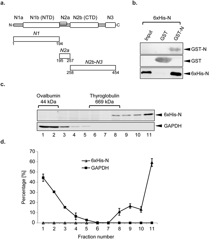
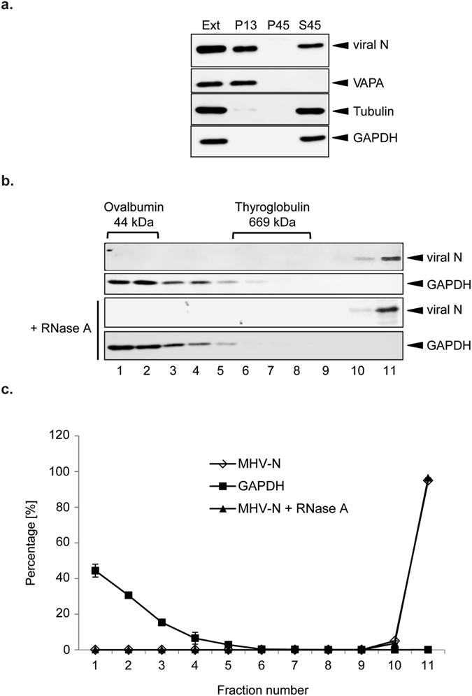
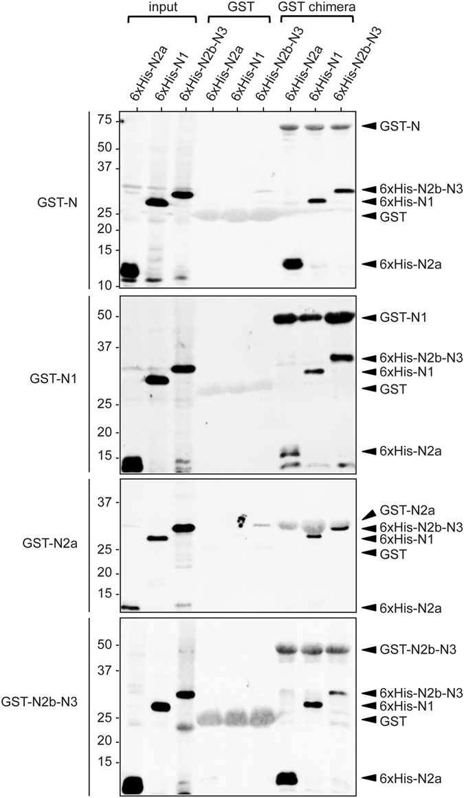
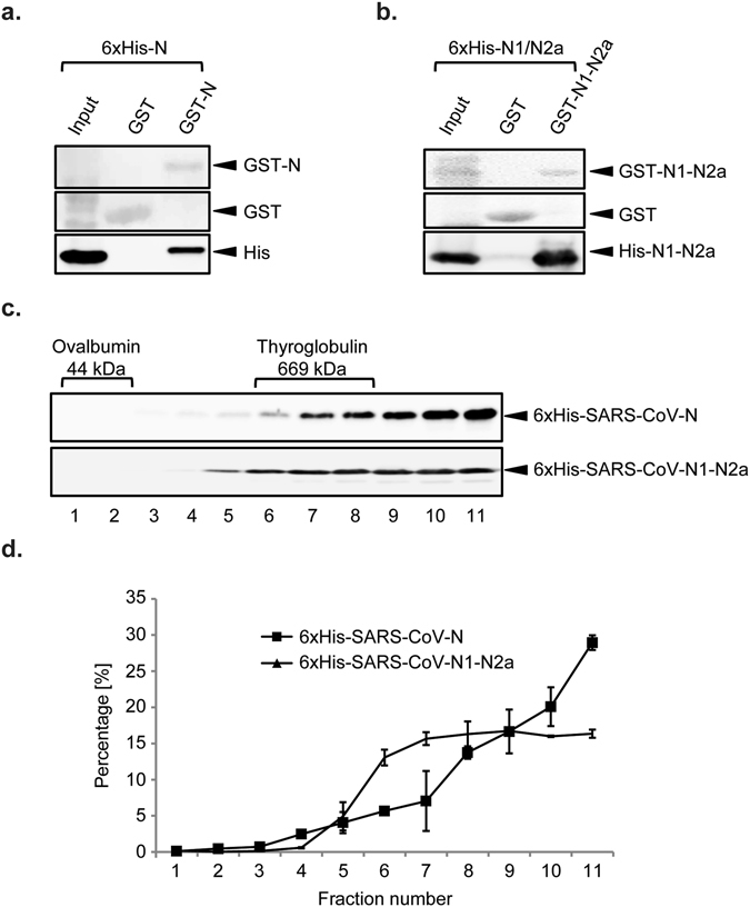
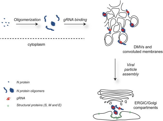
Coronaviruses (CoV) are enveloped viruses and rely on their nucleocapsid N protein to incorporate the positive-stranded genomic RNA into the virions. CoV N proteins form oligomers but the mechanism and relevance underlying their multimerization remain to be fully understood. Using in vitro pull-down experiments and density glycerol gradients, we found that at least 3 regions distributed over its entire length mediate the self-interaction of mouse hepatitis virus (MHV) and severe acute respiratory syndrome coronavirus (SARS-CoV) N protein. The fact that these regions can bind reciprocally between themselves provides a possible molecular basis for N protein oligomerization. Interestingly, cytoplasmic N molecules of MHV-infected cells constitutively assemble into oligomers through a process that does not require binding to genomic RNA. Based on our data, we propose a model where constitutive N protein oligomerization allows the optimal loading of the genomic viral RNA into a ribonucleoprotein complex via the presentation of multiple viral RNA binding motifs.


Conflict of interest statement

The authors declare that they have no competing interests.

Figures

Figure 1
Figure 1
Recombinant MHV N protein forms large oligomers. (a) Schematic structural organization of the MHV N protein and overview of the truncations generated in this study (modified from ref. 9). (b) Bacterial extract from E. coli expressing 6xHis-tagged MHV N protein (input) was incubated with immobilized GST or GST-N protein. Precipitated proteins were eluted in SDS-sample buffer and analyzed by western blot using an anti-His monoclonal antibody. Only part of the western blot images is shown in the figure. The GST and GST-N amount were assessed by staining the PVDF membrane with Ponceau Red. (c) Bacterial extract from E. coli expressing 6xHis-tagged MHV N protein was sedimented in a 5–20% glycerol gradient at 50,000 g for 75 min. Eleven fractions were collected and protein content analysed using antibodies against the 6xHis tag. Gradient and centrifugation conditions were assessed by sedimenting a LR7 cell extract and probing the collected fractions with a GAPDH antibody. Only part of the western blot images is shown in the figure. (d) Quantification of the immunoblots presented in panel (c) plus standard deviation (SD) (n = 3).
Figure 2
Figure 2
MHV N protein assembles in cytoplasmic oligomers over the course of an infection. (a) Cleared lysate from MHV-infected LR7 cells (Ext) was centrifuged at 15,000 × g for 10 min to obtain a pellet (P13) and a supernatant, which was subsequently centrifuged at 110,000 × g for 1 h to be separated into a pellet (P45) and a supernatant (S45). Equivalent amounts of each fraction were separated by SDS-PAGE and analysed by western blot using antibodies against MHV N protein, tubulin (cytoplasm), GAPDH (cytoplasm) and VAPA (endoplasmic reticulum). Only part of the western blot images is shown in the figure. (b) The S45 fraction obtained in panel (a) was mocked treated or treated with RNase A for 30 min on ice before sedimentation and analysed as in Fig. 1c. A260/280 ratios <0.1 was used to assess complete hydrolysis of the nucleic acids. Only part of the western blot images is shown in the figure. (c) Quantification of the immunoblots presented in panel (b) plus SD (n = 3).
Figure 3
Figure 3
Multiple domains mediate N protein oligomerization. Bacterial extracts from E. coli expressing the 6xHis-tagged N1, N2a and N2b-N3 truncations were incubated with immobilized GST, GST-N protein, GST-N1, GST-N2a and GST-N2b-N3. Precipitated proteins were eluted in SDS-sample buffer and analyzed by western blot using the anti-6xHis monoclonal antibody. A260/280 ratios <0.1 indicated the absence of nucleic acids in the samples.
Figure 4
Figure 4
SARS-CoV N protein is also forming oligomers. (a) Bacterial extract from E. coli expressing 6xHis-tagged SARS-CoV N protein (input) was incubated with immobilized GST or GST-SARS-CoV-N protein. Precipitated proteins were eluted in SDS-sample buffer and analyzed by western blot using an anti-His monoclonal antibody. The amounts of GST and GST-N were assessed by staining the PVDF membrane with Ponceau Red. A260/280 ratios <0.1 indicated the absence of nucleic acids in the samples. Only part of the western blot images is shown in the figure. (b) Bacterial extracts from E. coli expressing 6xHis-tagged SARS-CoV N1-N2a were processed and analyzed as in panel (a). A260/280 ratios <0.1 indicated absence of nucleic acids in the samples. Only part of the western blot images is shown in the figure. (c) Bacterial extracts of E. coli expressing 6xHis-tagged SARS N protein and N1-N2a truncation were sedimented in a 5–20% glycerol gradient at 50,000 g for 75 min. Eleven fractions were collected and protein content analyzed using antibodies against the 6xHis tag. Gradient and centrifugation conditions were assessed by sedimenting a LR7 cell extract and probing the collected fractions with a GAPDH antibody. Only part of the western blot images is shown in the figure. (d) Quantification of the immunoblots presented in panel (c) plus standard deviation (SD) (n = 3).
Figure 5
Figure 5
Models for the role of CoV N proteins over the course of an infection. After synthesis, CoV N proteins constitutively assemble into oligomers with loose or more compact intertwined filament shapes, which are recruited to the RTCs localized on double-membrane vesicles (DMVs) and convoluted membranes via their interaction with nsp3. At these replication platforms, newly synthesized gRNA is engaged by N protein oligomers, which co-operate with the rest of the structural proteins to form the viral particles at the ERGIC/Golgi compartments.

References

    1. Perlman S, Netland J. Coronaviruses post-SARS: update on replication and pathogenesis. Nat Rev Microbiol. 2009;7:439–50. doi: 10.1038/nrmicro2147. - DOI - PMC - PubMed
    1. Fehr AR, Perlman S. Coronaviruses: an overview of their replication and pathogenesis. Methods Mol Biol. 2015;1282:1–23. doi: 10.1007/978-1-4939-2438-7_1. - DOI - PMC - PubMed
    1. Cong Y, Ren X. Coronavirus entry and release in polarized epithelial cells: a review. Rev Med Virol. 2014;24:308–315. doi: 10.1002/rmv.1792. - DOI - PMC - PubMed
    1. Shehata MM, Gomaa MR, Ali MA, Kayali G. Middle East respiratory syndrome coronavirus: a comprehensive review. Front Med. 2016 - PMC - PubMed
    1. Chan HL, Tsui SK, Sung JJ. Coronavirus in severe acute respiratory syndrome (SARS) Trends Mol Med. 2003;9:323–325. doi: 10.1016/S1471-4914(03)00135-7. - DOI - PMC - PubMed

Publication types

Substances

LinkOut - more resources