

Tuberculosis - 'The great masquerader' presenting as a dumb-bell-shaped intradural extramedullary tumor in a 20-year-old female
- PMID: 28720995
- PMCID: PMC5498742
- DOI: 10.1016/j.jcot.2016.06.011
Tuberculosis - 'The great masquerader' presenting as a dumb-bell-shaped intradural extramedullary tumor in a 20-year-old female
Abstract
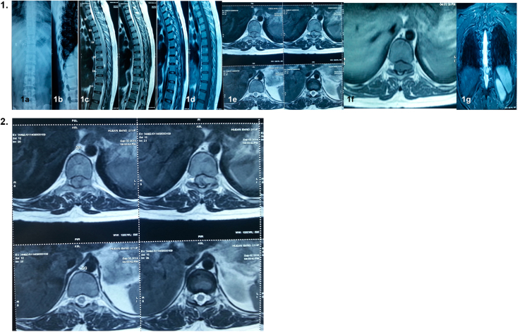
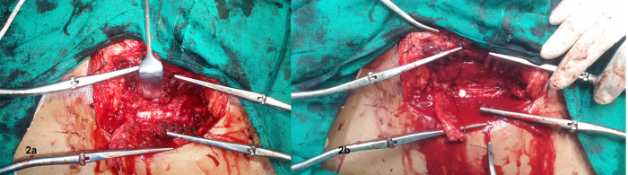
Tuberculosis has been known as the great masquerader for its varied presentations. We present an extraordinary case of a 20-year-old female who presented with paraparesis of two months. MRI showed an intradural, extramedullary dumb-bell-shaped, spinal cord tumor. With a provisional clinicoradiological diagnosis of benign nerve sheath tumor (schwannoma/neurofibroma), laminectomy was done. But after durotomy, frank pus was drained from the site of lesion and the laboratory investigations of the tissue and pus obtained proved it to be tubercular. This is a rare case reported in the literature where tuberculosis is mimicking as a dumb-bell-shaped, spinal cord tumor.
Keywords: Dumb-bell-shaped tumor; Spine; Tuberculosis.
Figures


References
-
- Ozawa H., Kokubun S., Aizawa T., Hoshikawa T., Kawahara C. Spinal dumb-bell tumours: an analysis of a series of 118 cases. J Neurosurg Spine. 2007;7(6):587–593. - PubMed
-
- Kumar S., Jain A.K., Dhammi I.K., Aggarwal A.N. Treatment of intraspinal tuberculoma. Clin Orthop Relat Res. 2007;460(July):62–66. - PubMed
-
- Luo L., Pino J. An intradural extramedullary tuberculoma of the spinal cord in a non-HIV-infected patient: case report and review of the literature. Lung. 2006;184(3):187–193. - PubMed
-
- Skendros P., Kamaria F., Kontopoulos V., Tsitouridis I., Sidiropoulos L. Intradural, extramedullary tuberculoma of the spinal cord as a complication of tuberculous meningitis. Infection. 2003;31(2):115–117. - PubMed
Publication types
LinkOut - more resources
Full Text Sources
Other Literature Sources
