

Mitochondria in oocyte aging: current understanding
- PMID: 28721182
- PMCID: PMC5506767
Mitochondria in oocyte aging: current understanding
Abstract
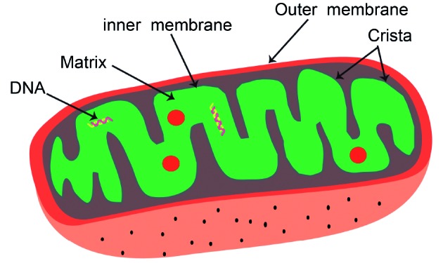
The oocyte is the largest cell found in multicellular organisms. Mitochondria, as the energy factories for cells, are found in high numbers in oocytes, as they provide the energy for oocyte maturation, fertilization, and embryo formation via oxidative phosphorylation. Failure of assisted reproduction is mainly attributed to oocyte aging and increased aneuploidy. As the most numerous organelle in the oocyte, the mitochondrion has been confirmed as a crucial player in the process of oocyte aging, which is highly influenced by mitochondrion dysfunction. Every mitochondrion contains one or more mitochondrial DNA (mtDNA) molecule, which, at about 16.5 KD in length, encodes 13 proteins. In this review, we discuss the function of mitochondria and the relationship between mtDNA and oocyte aging. We also discuss technologies that aim to enhance oocyte developmental potential and delay ovarian aging.
Keywords: Oocyte; aging; embryo; mitochondria; mtDNA.
Figures
References
-
- Aiken CE, Cindrova-Davies T, Johnson MH. Variations in mouse mitochondrial DNA copy number from fertilization to birth are associated with oxidative stress. Reprod Biomed Online. 2008;17(6):806–813. - PubMed
-
- Barritt JA, Cohen J, Brenner CA. Mitochondrial DNA point mutation in human oocytes is associated with maternal age. Reprod Biomed Online. 2000;1(3):96–100. - PubMed
-
- Battaglia DE, Goodwin P, Klein NA, et al. Influence of maternal age on meiotic spindle assembly in oocytes from naturally cycling women. Hum Reprod. 1996;11(10):2217–2222. - PubMed
-
- Bauer G. Targeting extracellular ROS signaling of tumor cells. Anticancer Res. 2014;34(4):1467–1482. - PubMed
Publication types
LinkOut - more resources
Full Text Sources
Research Materials