

A glimpse at the aging eye
- PMID: 28721262
- PMCID: PMC5515005
- DOI: 10.1038/npjamd.2016.3
A glimpse at the aging eye
Abstract
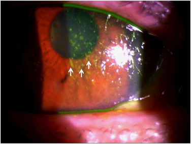
Extensive investigations have demonstrated that organismal aging is associated with tissue dysfunction in many organs. The eye is no exception to this rule. Under healthy conditions, the eye is designed like an advanced camera with the central role of translating light from the external world into a coherent neural signal that can be transmitted to the brain for processing into a precise visual image. This complex process requires precisely maintained machinery. At the front of the eye, the transparency of both the cornea and the lens are crucial to allow passage of photons to the light-sensitive portion of the eye. Similarly, the highly organized structure of the retina located at the back of the eye is indispensable to allow for effective signal transduction and efficient signal transmission. Aging affects ocular structures in various ways, and these sequelae have been well defined as distinct clinical entities. In many instances, aging leads to ocular tissue dysfunction and disease. Nonetheless, despite clear evidence that age-associated visual impairment has significant psychosocial consequences, current treatment paradigms for many of these conditions are inadequate. In addition, strategies to decelerate or reverse age-associated deterioration in ocular function are still in their infancy. This review focuses on the cellular and molecular pathophysiology of the aging eye. Ultimately, we hope that a refined understanding of the aging eye can guide targeted therapies against cellular aging and disease.
Conflict of interest statement
The authors declare no conflict of interest.
Figures
References
-
- Miratashi, S. A. M. , Besharati, M. R. , Manaviat, M. R. , Rastegar, A. & Shoja, M. R. Vitamin C concentration of aqueous humour and plasma in patients with senile cataract. Asian J. Ophthalmol. 6, 6–9 (2004).
-
- Agte, V. & Tarwadi, K. The importance of nutrition in the prevention of ocular disease with special reference to cataract. Ophthalmic Res. 44, 166–172 (2010). - PubMed
-
- Gipson, I. K. Age-related changes and diseases of the ocular surface and cornea. Invest. Ophthalmol. Vis. Sci. 54, ORSF48–ORSF53 (2013). - PubMed
Publication types
Grants and funding
LinkOut - more resources
Full Text Sources
Other Literature Sources
