. 2017 Sep 8;292(36):14885-14901.
doi: 10.1074/jbc.M117.804344. Epub 2017 Jul 19.

Heterodimers of the transcriptional factors NFATc3 and FosB mediate tissue factor expression for 15(S)-hydroxyeicosatetraenoic acid-induced monocyte trafficking

Affiliations

Heterodimers of the transcriptional factors NFATc3 and FosB mediate tissue factor expression for 15(S)-hydroxyeicosatetraenoic acid-induced monocyte trafficking

Sivareddy Kotla et al. J Biol Chem. .

Erratum in

Abstract

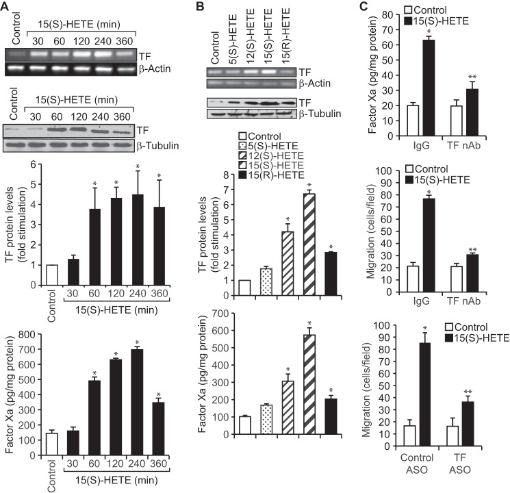
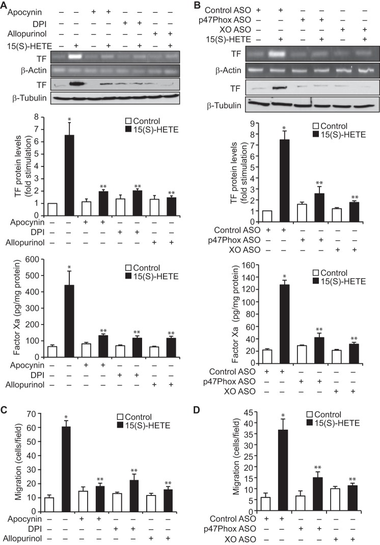
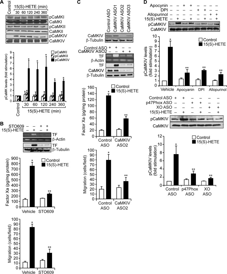
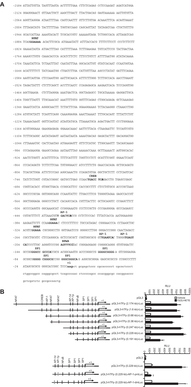
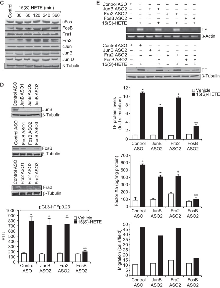
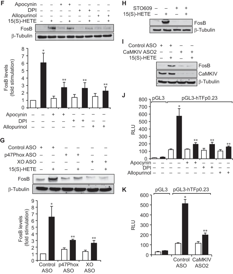
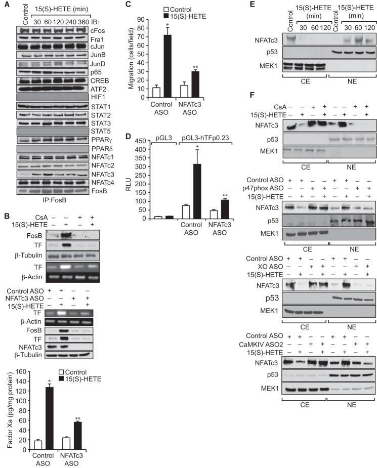
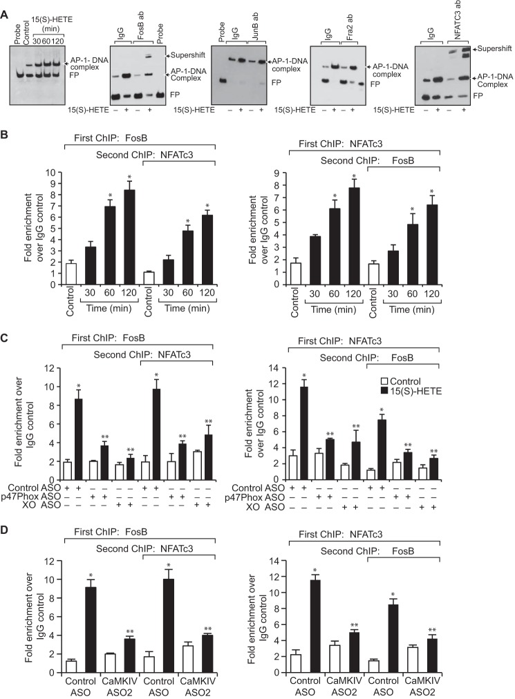
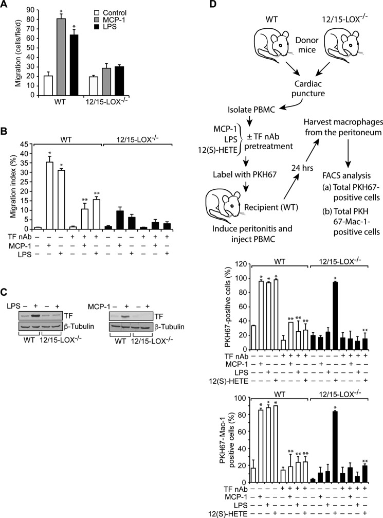
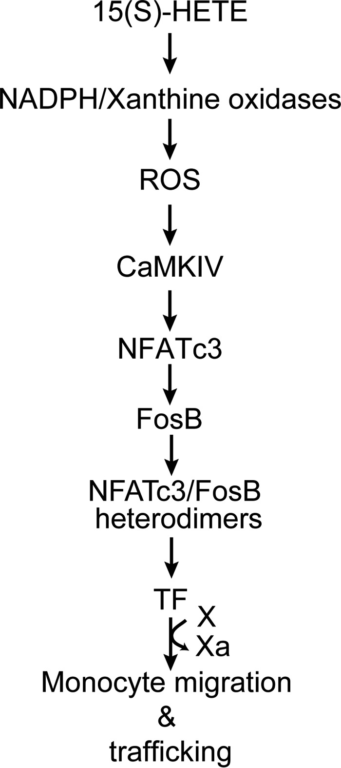
Tissue factor (TF) is expressed in vascular and nonvascular tissues and functions in several pathways, including embryonic development, inflammation, and cell migration. Many risk factors for atherosclerosis, including hypertension, diabetes, obesity, and smoking, increase TF expression. To better understand the TF-related mechanisms in atherosclerosis, here we investigated the role of 12/15-lipoxygenase (12/15-LOX) in TF expression. 15(S)-hydroxyeicosatetraenoic acid (15(S)-HETE), the major product of human 15-LOXs 1 and 2, induced TF expression and activity in a time-dependent manner in the human monocytic cell line THP1. Moreover, TF suppression with neutralizing antibodies blocked 15(S)-HETE-induced monocyte migration. We also found that NADPH- and xanthine oxidase-dependent reactive oxygen species (ROS) production, calcium/calmodulin-dependent protein kinase IV (CaMKIV) activation, and interactions between nuclear factor of activated T cells 3 (NFATc3) and FosB proto-oncogene, AP-1 transcription factor subunit (FosB) are involved in 15(S)-HETE-induced TF expression. Interestingly, NFATc3 first induced the expression of its interaction partner FosB before forming the heterodimeric NFATc3-FosB transcription factor complex, which bound the proximal AP-1 site in the TF gene promoter and activated TF expression. We also observed that macrophages from 12/15-LOX-/- mice exhibit diminished migratory response to monocyte chemotactic protein 1 (MCP-1) and lipopolysaccharide compared with WT mouse macrophages. Similarly, compared with WT macrophages, monocytes from 12/15-LOX-/- mice displayed diminished trafficking, which was rescued by prior treatment with 12(S)-HETE, in a peritonitis model. These observations indicate that 15(S)-HETE-induced monocyte/macrophage migration and trafficking require ROS-mediated CaMKIV activation leading to formation of NFATc3 and FosB heterodimer, which binds and activates the TF promoter.

Keywords: gene expression; migration; monocyte; signal transduction; trafficking.


Conflict of interest statement

The authors declare that they have no conflicts of interest with the contents of this article

Figures

Figure 1.
Figure 1.
TF mediates 15(S)-HETE–induced THP1 cell migration. A, quiescent THP1 cells were treated with vehicle or 15(S)-HETE (0.1 μm) for the indicated time periods and either RNA was isolated or cell extracts were prepared. The RNA and cell extracts were analyzed by RT-PCR and Western blotting for TF mRNA and protein levels using its specific primers and antibodies, respectively. TF activity in cell extracts was measured using a kit as described in “Materials and methods.” B, quiescent THP1 cells were treated with vehicle or the indicated HETE (0.1 μm) for 2 h, and TF mRNA, protein, and activity were measured as described in A. The mRNA and protein levels in A and B were normalized to β-actin mRNA and β-tubulin protein levels, respectively. C, quiescent THP1 cells were incubated with IgG or TF neutralizing antibodies (anti-human TF Ab-D3, 5 μg/ml) for 30 min, treated with vehicle or 15(S)-HETE (0.1 μm) for either 2 h and TF activity was measured or 8 h and cell migration was measured. Cells were also transfected with control or TF ASOs (100 nm), quiesced, and treated with vehicle or 15(S)-HETE, and migration was measured. The bar graphs represent mean ± S.D. values of three experiments. *, p < 0.01 versus vehicle control or control ASO; **, p < 0.01 versus 15(S)-HETE, TF, or control ASO + 15(S)-HETE.
Figure 2.
Figure 2.
NADPH oxidase and xanthine oxidase-dependent ROS production is required for 15(S)-HETE–induced TF expression and its activity in mediating THP1 cell migration. A, quiescent THP1 cells are treated with vehicle or 15(S)-HETE (0.1 μm) in the presence and absence of apocynin (100 μm), DPI (10 μm), or allopurinol (100 μm) for 2 h, and TF expression (mRNA and protein levels) and its activity were measured. B, cells were transfected with control, p47Phox, or xanthine oxidase ASOs, quiesced, and treated with vehicle or 15(S)-HETE (0.1 μm) for 2 h, and TF expression (mRNA and protein levels) and its activity were measured. C and D, all the conditions were the same as in A or B except that cells were treated with vehicle or 15(S)-HETE (0.1 μm) for 8 h, and cell migration was measured. The mRNA and protein levels in A and B were normalized to β-actin mRNA and β-tubulin protein levels, respectively. The bar graphs represent mean ± S.D. of three experiments. *, p < 0.01 versus vehicle control or control ASO; **, p < 0.01 versus 15(S)-HETE or control ASO + 15(S)-HETE.
Figure 3.
Figure 3.
15(S)-HETE–induced TF expression and its activity require CaMKIV activation. A, quiescent THP1 cells treated with vehicle or 15(S)-HETE (0.1 μm) for the indicated time periods, and cell extracts were prepared. Equal amounts of protein from control and each treatment were analyzed by Western blotting for the phosphorylation of the indicated CaMKs using their specific antibodies and normalized to their total levels. B, quiescent THP1 cells were treated with vehicle or 15(S)-HETE (0.1 μm) in the presence and absence of STO609 (10 μm) either for 2 h and TF expression (mRNA and protein levels) and its activity were measured, or for 8 h and cell migration was measured. C, upper panel, THP1 cells were transfected with control or the indicated ASOs of CaMKIV, and 48 h later cell extracts were prepared and analyzed by Western blotting for CaMKIV levels using its specific antibodies and normalized to β-tubulin levels. Lower panels, THP1 cells that were transfected with control or the indicated CaMKIV ASO and quiesced were treated with vehicle or 15(S)-HETE (0.1 μm) either for 2 h and analyzed for TF expression (mRNA and protein levels) and its activity or for 8 h and cell migration was measured. D, quiescent THP1 cells were treated with vehicle or 15(S)-HETE (0.1 μm) in the presence and absence of apocynin (100 μm), DPI (10 μm), or allopurinol (100 μm) for 1 h or transfected with control, p47Phox, or xanthine oxidase ASO and quiesced before subjecting to treatment with vehicle or 15(S)-HETE (0.1 μm) for 1 h and cell extracts were prepared. Equal amounts of protein from control and each treatment were analyzed by Western blotting for pCaMKIV levels using its specific antibodies and normalized to its total levels. The mRNA and protein levels in B and C were normalized to β-actin mRNA and β-tubulin protein levels. The bar graphs represent mean ± S.D. values of three independent experiments. *, p < 0.01 versus vehicle control or control ASO; **, p < 0.01 versus vehicle control + 15(S)-HETE or control ASO + 15(S)-HETE.
Figure 4.
Figure 4.
FosB mediates 15(S)-HETE–induced TF promoter activity. A, TF promoter encompassing from −2174 nt to +116 nt was cloned and sequenced. B, TF promoter-luciferase constructs with serial 5′ truncations or site-directed insertion of mutations into the indicated AP-1 site in the pGL3-hTFp (0.23 kb)-Luc construct were made, transfected into THP1 cells, quiesced, treated with vehicle or 15(S)-HETE (0.1 μm) for 6 h and their activities were measured. AP-1p and AP-1d represent the proximal and distal AP-1–binding sites located at −205, and −217 nt, respectively. C, quiescent THP1 cells were treated with vehicle or 15(S)-HETE (0.1 μm) for the indicated time periods, and the cell extracts were prepared and analyzed by Western blotting for the indicated proteins using their specific antibodies. D, THP1 cells were transfected with control or the indicated JunB, FosB, or Fra2 ASOs and 48 h later cell extracts were prepared and analyzed by Western blotting for JunB, FosB, or Fra2 levels using their specific antibodies and normalized to β-tubulin. THP1 cells were co-transfected with TF promoter-luciferase construct in combination with or without control, JunB, FosB, or Fra2 ASOs, quiesced, treated with vehicle or 15(S)-HETE (0.1 μm) for 6 h, and analyzed for luciferase activity. E, THP1 cells that were transfected with control, JunB, FosB, or Fra2 ASOs and quiesced were treated with vehicle or 15(S)-HETE (0.1 μm) either for 2 h and analyzed for TF expression (mRNA and proteins levels) and its activity or for 8 h and cell migration was measured. F–I, the role of NADPH oxidase, xanthine oxidase, and CaMKIV in 15(S)-HETE–induced FosB expression was determined using pharmacological and ASO approaches as described in Fig. 2, A and B, and Fig. 3, B and C, respectively. The blots in F–I are reprobed with β-tubulin for normalization. J, THP1 cells were transfected with empty vector or pGL3-hTFp0.23-Luc construct, quiesced, treated with vehicle or 15(S)-HETE (0.1 μm) in the presence and absence of apocynin (100 μm), DPI (10 μm), or allopurinol (100 μm) for 6 h, and cell extracts were prepared and analyzed for luciferase activity. K, THP1 cells were co-transfected with empty vector or pGL3-hTFp0.23-Luc in combination with and without control or CaMKIV ASO, quiesced, treated with vehicle or 15(S)-HETE (0.1 μm) for 6 h, and cell extracts were prepared and analyzed for luciferase activity. *, p < 0.01 versus vehicle control or control ASO or empty vector; **, p < 0.01 versus vehicle control + 15(S)-HETE or control ASO + 15(S)-HETE or pGL3-hTF + 15(S)-HETE.
Figure 4.
Figure 4.
FosB mediates 15(S)-HETE–induced TF promoter activity. A, TF promoter encompassing from −2174 nt to +116 nt was cloned and sequenced. B, TF promoter-luciferase constructs with serial 5′ truncations or site-directed insertion of mutations into the indicated AP-1 site in the pGL3-hTFp (0.23 kb)-Luc construct were made, transfected into THP1 cells, quiesced, treated with vehicle or 15(S)-HETE (0.1 μm) for 6 h and their activities were measured. AP-1p and AP-1d represent the proximal and distal AP-1–binding sites located at −205, and −217 nt, respectively. C, quiescent THP1 cells were treated with vehicle or 15(S)-HETE (0.1 μm) for the indicated time periods, and the cell extracts were prepared and analyzed by Western blotting for the indicated proteins using their specific antibodies. D, THP1 cells were transfected with control or the indicated JunB, FosB, or Fra2 ASOs and 48 h later cell extracts were prepared and analyzed by Western blotting for JunB, FosB, or Fra2 levels using their specific antibodies and normalized to β-tubulin. THP1 cells were co-transfected with TF promoter-luciferase construct in combination with or without control, JunB, FosB, or Fra2 ASOs, quiesced, treated with vehicle or 15(S)-HETE (0.1 μm) for 6 h, and analyzed for luciferase activity. E, THP1 cells that were transfected with control, JunB, FosB, or Fra2 ASOs and quiesced were treated with vehicle or 15(S)-HETE (0.1 μm) either for 2 h and analyzed for TF expression (mRNA and proteins levels) and its activity or for 8 h and cell migration was measured. F–I, the role of NADPH oxidase, xanthine oxidase, and CaMKIV in 15(S)-HETE–induced FosB expression was determined using pharmacological and ASO approaches as described in Fig. 2, A and B, and Fig. 3, B and C, respectively. The blots in F–I are reprobed with β-tubulin for normalization. J, THP1 cells were transfected with empty vector or pGL3-hTFp0.23-Luc construct, quiesced, treated with vehicle or 15(S)-HETE (0.1 μm) in the presence and absence of apocynin (100 μm), DPI (10 μm), or allopurinol (100 μm) for 6 h, and cell extracts were prepared and analyzed for luciferase activity. K, THP1 cells were co-transfected with empty vector or pGL3-hTFp0.23-Luc in combination with and without control or CaMKIV ASO, quiesced, treated with vehicle or 15(S)-HETE (0.1 μm) for 6 h, and cell extracts were prepared and analyzed for luciferase activity. *, p < 0.01 versus vehicle control or control ASO or empty vector; **, p < 0.01 versus vehicle control + 15(S)-HETE or control ASO + 15(S)-HETE or pGL3-hTF + 15(S)-HETE.
Figure 5.
Figure 5.
NFATc3 mediates 15(S)-HETE–induced TF expression and activity. A, equal amounts of protein from control and the indicated time periods of 15(S)-HETE (0.1 μm)–treated cells were immunoprecipitated with anti-FosB antibodies, and the immunocomplexes were analyzed by Western blotting for the indicated proteins using their specific antibodies followed by normalization to FosB. B, quiescent THP1 cells were treated with vehicle or 15(S)-HETE (0.1 μm) in the presence or absence of CsA (10 μm) for 2 h and analyzed for TF expression (mRNA and protein levels) and its activity. All the conditions were the same as in the upper panel except that cells were transfected with control or NFATc3 ASOs and quiesced before subjecting to treatment with and without 15(S)-HETE. C, THP1 cells were transfected with control or NFATc3 ASOs, quiesced, and subjected to 15(S)-HETE (0.1 μm)–induced migration. D, THP1 cells were co-transfected with empty vector or pGL3-hTFp in combination with and without control or NFTAc3 ASOs, quiesced, treated with vehicle or 15(S)-HETE (0.1 μm) for 6 h; cell extracts were prepared and analyzed for luciferase activity. E, quiesced THP1 cells were treated with vehicle or 15(S)-HETE (0.1 μm) for the indicated time periods, and the cytoplasmic extracts (CE) and nuclear extracts (NE) were prepared and analyzed by Western blotting for the indicated proteins using their specific antibodies. F, THP1 cells were transfected with control, p47phox, xanthine oxidase, or CaMKIV ASOs, quiesced, treated with vehicle or 15(S)-HETE (0.1 μm) for the indicated time periods, and the CE and NE were prepared and analyzed by Western blotting for the indicated proteins using their specific antibodies. *, p < 0.01 versus control ASO; **, p < 0.01 versus control ASO + 15(S)-HETE.
Figure 6.
Figure 6.
NFATc3 and FosB interact with each other and bind to AP-1 site in the TF promoter. A, nuclear extracts of control and the indicated time periods or 2 h of 15(S)-HETE (0.1 μm)–treated cells were analyzed for AP-1 and DNA activity and for the presence of FosB, JunB, Fra2, or NFATc3 in the AP-1-DNA complexes by EMSA and supershift EMSA, respectively, using AP-1–binding site at −205 nt in the TF promoter as a biotin-labeled probe. B, THP1 cells were treated with vehicle or 15(S)-HETE (0.1 μm) for the indicated time periods and subjected to ChIP and re-ChIP assay of TF promoter using anti-FosB or anti-NFATc3 antibodies. C and D, THP1 cells were transfected with control, p47phox, xanthine oxidase, or CaMKIV ASOs, quiesced, treated with vehicle or 15(S)-HETE (0.1 μm) for 2 h and subjected to ChIP and re-ChIP assays for FosB and NFATc3 binding to TF promoter as described in B. *, p < 0.01 versus control ASO; **, p < 0.01 versus control ASO + 15(S)-HETE. FP, free probe.
Figure 7.
Figure 7.
12/15-LOX/15(S)-HETE is required for monocyte/macrophage trafficking. A, peritoneal macrophages from WT and 12/15-LOX−/− mice were isolated and subjected to MCP-1 (20 ng/ml) and LPS (100 ng/ml)–induced migration. B, in vivo migration of peritoneal macrophages induced by MCP-1 or LPS in presence and absence of TF neutralizing antibodies (anti-mouse TF Ab-1H1) was performed using macrophage efflux model as described in “Materials and methods.” C, peritoneal macrophages from WT and 12/15-LOX−/− mice were isolated and treated with and without MCP-1 (20 ng/ml) or LPS (100 ng/ml) for 2 h. Cell extracts were prepared and analyzed by Western blotting for TF levels using its specific antibodies, and the blot was normalized for β-tubulin. D, PBMC from WT and 12/15-LOX−/− mice were isolated, incubated with TF nAb for 1 h at room temperature, treated with and without MCP-1, LPS, or 12(S)-HETE for 2 h, labeled with PKH67, and injected via tail vein into peritonitis recipient WT mice. After 24 h, peritoneal cells were harvested and the number of PKH67-positive as well as PKH67+Mac-1–positive cells were quantified by flow cytometry. *, p < 0.01 versus vehicle control; **, p < 0.01 versus WT + MCP-1 or WT + LPS or WT + 12(S)-HETE or 12/15-LOX−/− + 12(S)-HETE.
Figure 8.
Figure 8.
Schematic diagram depicting the potential mechanism of 15(S)-HETE–induced TF expression and monocyte migration/trafficking.

References

    1. Ruf W., and Edgington T. S. (1994) Structural biology of tissue factor, the initiator of thrombogenesis in vivo. FASEB J. 8, 385–390 - PubMed
    1. Pawlinski R., Fernandes A., Kehrle B., Pedersen B., Parry G., Erlich J., Pyo R., Gutstein D., Zhang J., Castellino F., Melis E., Carmeliet P., Baretton G., Luther T., Taubman M., Rosen E., and Mackman N. (2002) Tissue factor deficiency causes cardiac fibrosis and left ventricular dysfunction. Proc. Natl. Acad. Sci. U.S.A. 99, 15333–15338 - PMC - PubMed
    1. Nishibe T., Parry G., Ishida A., Aziz S., Murray J., Patel Y., Rahman S., Strand K., Saito K., Saito Y., Hammond W. P., Savidge G. F., Mackman N., and Wijelath E. S. (2001) Oncostatin M promotes biphasic tissue factor expression in smooth muscle cells: Evidence for Erk-1/2 activation. Blood 97, 692–699 - PubMed
    1. Parry G. C., and Mackman N. (2000) Mouse embryogenesis requires the tissue factor extracellular domain but not the cytoplasmic domain. J. Clin. Invest. 105, 1547–1554 - PMC - PubMed
    1. Chen J., Kasper M., Heck T., Nakagawa K., Humpert P. M., Bai L., Wu G., Zhang Y., Luther T., Andrassy M., Schiekofer S., Hamann A., Morcos M., Chen B., Stern D. M., Nawroth P. P., and Bierhaus A. (2005) Tissue factor as a link between wounding and tissue repair. Diabetes 54, 2143–2154 - PubMed

MeSH terms

LinkOut - more resources