. 2017 Oct;28(10):3076-3088.
doi: 10.1681/ASN.2016050598. Epub 2017 Jul 19.

Histopathology of Veins Obtained at Hemodialysis Arteriovenous Fistula Creation Surgery

Collaborators, Affiliations

Histopathology of Veins Obtained at Hemodialysis Arteriovenous Fistula Creation Surgery

Charles E Alpers et al. J Am Soc Nephrol. 2017 Oct.

Abstract

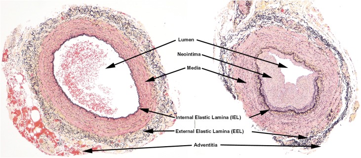
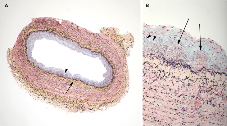
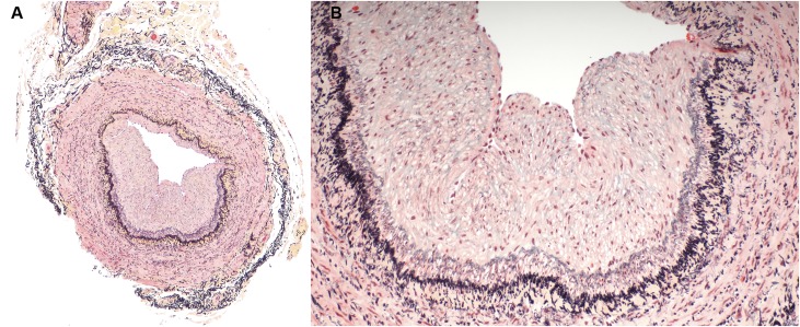
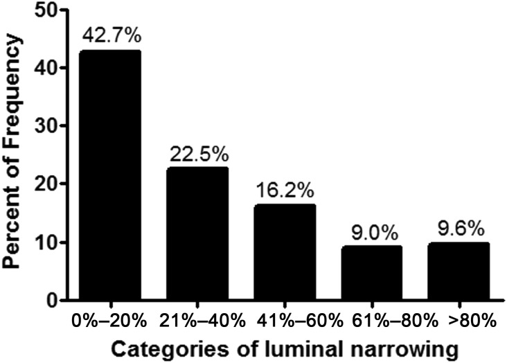
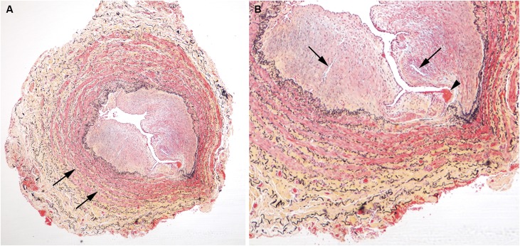
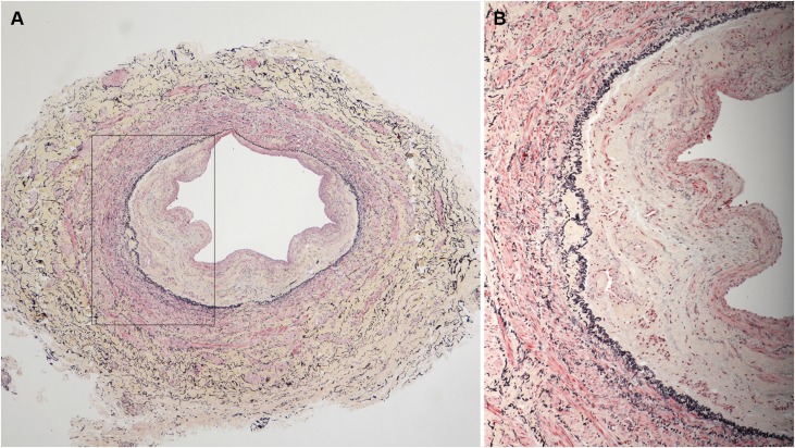
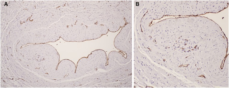
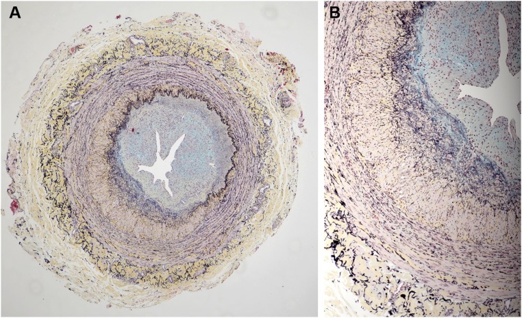
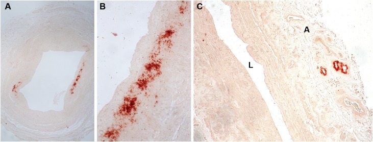
Stenosis from venous neointimal hyperplasia is common in native arteriovenous fistulas (AVFs). However, the preexisting histologic characteristics of veins at fistula creation, and associations thereof with baseline patient factors, have not been well characterized. In this study, we conducted histologic analysis of a segment of the vein used for anastomosis creation, obtained during AVF creation from 554 of the 602 participants in the multicenter Hemodialysis Fistula Maturation Cohort Study. We quantified intimal and medial areas and lengths of the internal and external elastic lamina by morphometry and assessed venous wall cells by immunohistochemistry, extracellular matrix with Movat stain, and calcium deposition by alizarin red stain. We also studied a representative subset of veins for markers of monocyte/macrophage content, cell proliferation, apoptosis, and neoangiogenesis. Neointima occupied >20% of the lumen in 57% of fully circumferential vein samples, and neointimal hyperplasia associated positively with age and inversely with black race. The neointima was usually irregularly thickened, sometimes concentric, and contained α-smooth muscle actin-expressing cells of smooth muscle or myofibroblast origin. Proteoglycans admixed with lesser amounts of collagen constituted the predominant matrix in the neointima. In 82% of vein samples, the media of vessel walls contained large aggregates of collagen. A minority of veins expressed markers of inflammation, cell proliferation, cell death, calcification, or neoangiogenesis. In conclusion, we observed preexisting abnormalities, including neointimal hyperplasia and prominent accumulation of extracellular matrix, in veins used for AVF creation from a substantial proportion of this cohort.

Keywords: arteriovenous access; arteriovenous fistula; dialysis access; pathology.


Figures

Figure 1.
Figure 1.
Illustration of characteristic landmarks of both normal veins and abnormal veins. There is a neointima and accumulation of medial collagen (yellow staining) adjacent to the IEL in the abnormal vein (right). In the abnormal veins, the indicated landmarks were measured morphometrically to determine the extent of luminal compromise by neointima (Movat pentachrome stain).
Figure 2.
Figure 2.
Vein with concentric neointima formation. A lower power image is on the left (A) and a higher power image of a section of the same vein is shown on the right (B). (A) The neointima contains large amounts of blue staining material (proteoglycans, arrowhead), whereas there is prominent concentric accumulation of collagen (yellow stain, long arrow) in the media immediately adjacent to the IEL. No significant inflammatory infiltrates are identified. (B) Discrete microdomains of admixed collagen (yellow) and proteoglycans (blue), indicated by arrows, were observed within the neointima. Some microdomains appeared to have a component of neoangiogenesis (arrowheads). (Movat pentachrome stain)
Figure 3.
Figure 3.
Abnormal vein with eccentric neointima formation, stained with Movat pentachrome. (A) is a low power image and (B) is a higher power image of the vein section shown on the right in Figure 1. Less proteoglycans (blue staining) are noted in the neointima of this sample and less collagen (yellow staining) is noted in the vessel wall. Inflammatory infiltrates are not present in any of the layers of the vessel wall.
Figure 4.
Figure 4.
Graphic depiction of the extent of luminal narrowing by formation of a neointima in the 365 veins undergoing morphometric analysis. The majority of veins (57%) show 20% or greater encroachment of the lumen area by neointima. Nearly 10% of veins showed severe luminal narrowing of 80% or more.
Figure 5.
Figure 5.
Example of vein, stained with Movat pentachrome, showing highest degree of luminal occlusion by a neointima. (A) This vein shows accumulations of collagen (yellow-stained matrix, arrows) dispersed throughout the media. (B) High-power view of vein in (A) shows a small thrombus (arrowhead) at the neointimal surface. Several small vessels within a more cellular region of the neointima are present (arrows). The IEL (black stain) is disrupted in the portion of the vein near the thrombus.
Figure 6.
Figure 6.
A vein sample, stained with Movat pentachrome, with a very eccentric neointimal layer. A lower power image is on the left (A) and a higher power image (B) of a section of the same vein is shown on the right that was obtained from the vessel area indicated by the black outline in (A). This sample had a large component of collagen (yellow staining) in the neointimal and adventitial layers with very little proteoglycan content (blue staining).
Figure 7.
Figure 7.
Immunostaining with anti-CD31 antibody to detect endothelial cells. Rust color indicates positive immunostaining. A lower power image is on the left (A) and a higher power image of a section of the same vein is shown on the right (B). Endothelial cells lining the vein lumen are highlighted. Positive immunostaining of microvessels in the neointima and adventitia is visible in (B), suggesting neoangiogenesis in these regions.
Figure 8.
Figure 8.
A vein sample, stained with Movat pentachrome, with a very eccentric neointimal layer. A lower power image is on the left (A) and a higher power image of a section of the same vein is shown on the right (B). This sample had large aggregates of collagen (yellow staining) in the media adjacent to the IEL and in the adventitia, with large proteoglycan content (blue staining) in the neointima.
Figure 9.
Figure 9.
Examples of calcification in vein detected by alizarin red S reagent. A lower power image (A) shows discrete areas of calcium accumulation (dark red). A higher power image of the calcium deposits in (A) is shown in (B). In (C), another vein sample had calcium deposits confined to neovessels in the adventitia (A). Calcification was typically detected in the medial and adventitial layers of veins with little accumulation of calcium within neointimas. A, adventitia; L, vein lumen.

Comment in

References

    1. Schinstock CA, Albright RC, Williams AW, Dillon JJ, Bergstralh EJ, Jenson BM, McCarthy JT, Nath KA: Outcomes of arteriovenous fistula creation after the fistula first initiative. Clin J Am Soc Nephrol 6: 1996–2002, 2011 - PMC - PubMed
    1. Dixon BS: Why don’t fistulas mature? Kidney Int 70: 1413–1422, 2006 - PubMed
    1. Allon M, Litovsky S, Young CJ, Deierhoi MH, Goodman J, Hanaway M, Lockhart ME, Robbin ML: Medial fibrosis, vascular calcification, intimal hyperplasia, and arteriovenous fistula maturation. Am J Kidney Dis 58: 437–443, 2011 - PMC - PubMed
    1. Allon M, Litovsky S, Young CJ, Deierhoi MH, Goodman J, Hanaway M, Lockhart ME, Robbin ML: Correlation of pre-existing vascular pathology with arteriovenous graft outcomes in hemodialysis patients. Am J Kidney Dis 62: 1122–1129, 2013 - PMC - PubMed
    1. Allon M, Robbin ML, Young CJ, Deierhoi MH, Goodman J, Hanaway M, Lockhart ME, Litovsky S: Preoperative venous intimal hyperplasia, postoperative arteriovenous fistula stenosis, and clinical fistula outcomes. Clin J Am Soc Nephrol 8: 1750–1755, 2013 - PMC - PubMed

LinkOut - more resources