. 2017 Jul 20;2(14):e94382.
doi: 10.1172/jci.insight.94382.

Pediatric and adult dilated cardiomyopathy represent distinct pathological entities

Affiliations

Pediatric and adult dilated cardiomyopathy represent distinct pathological entities

Meghna D Patel et al. JCI Insight. .

Abstract

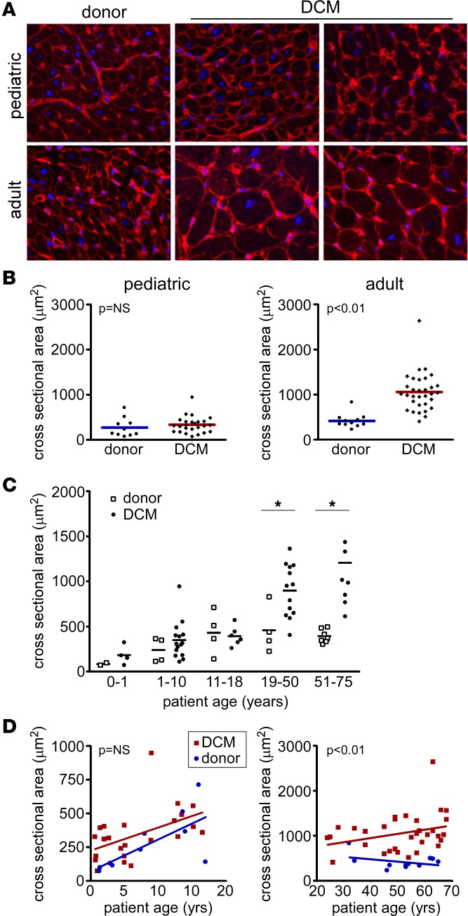
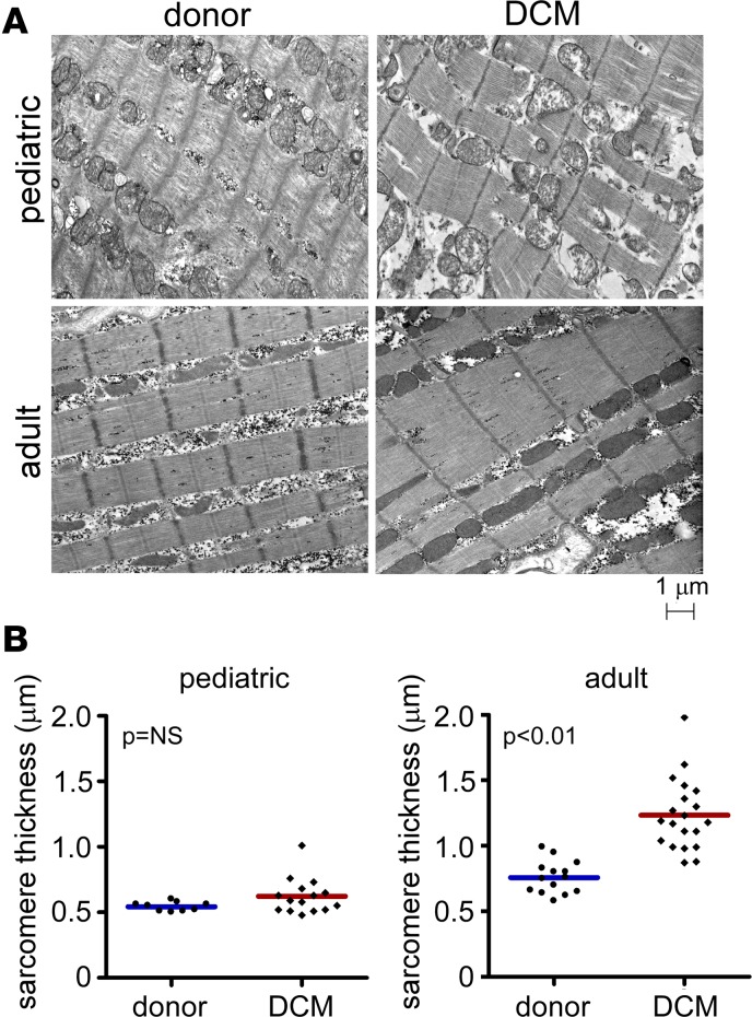
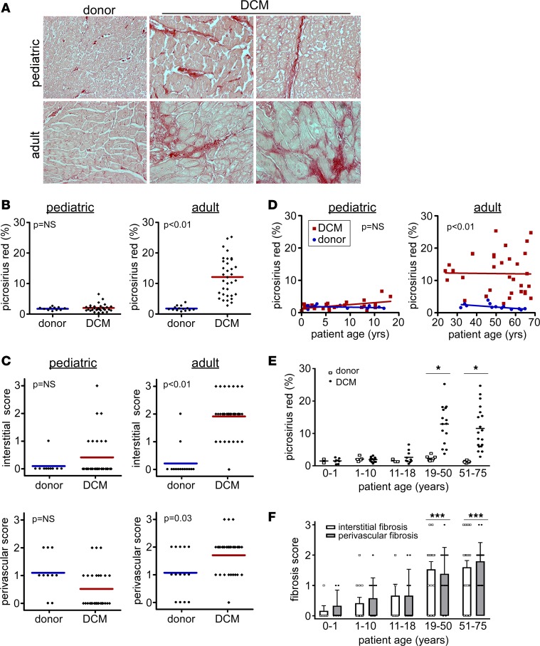
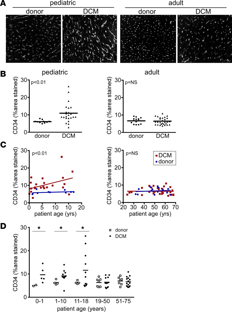
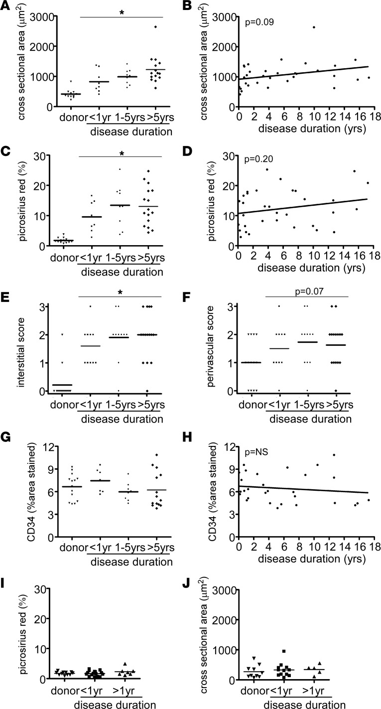
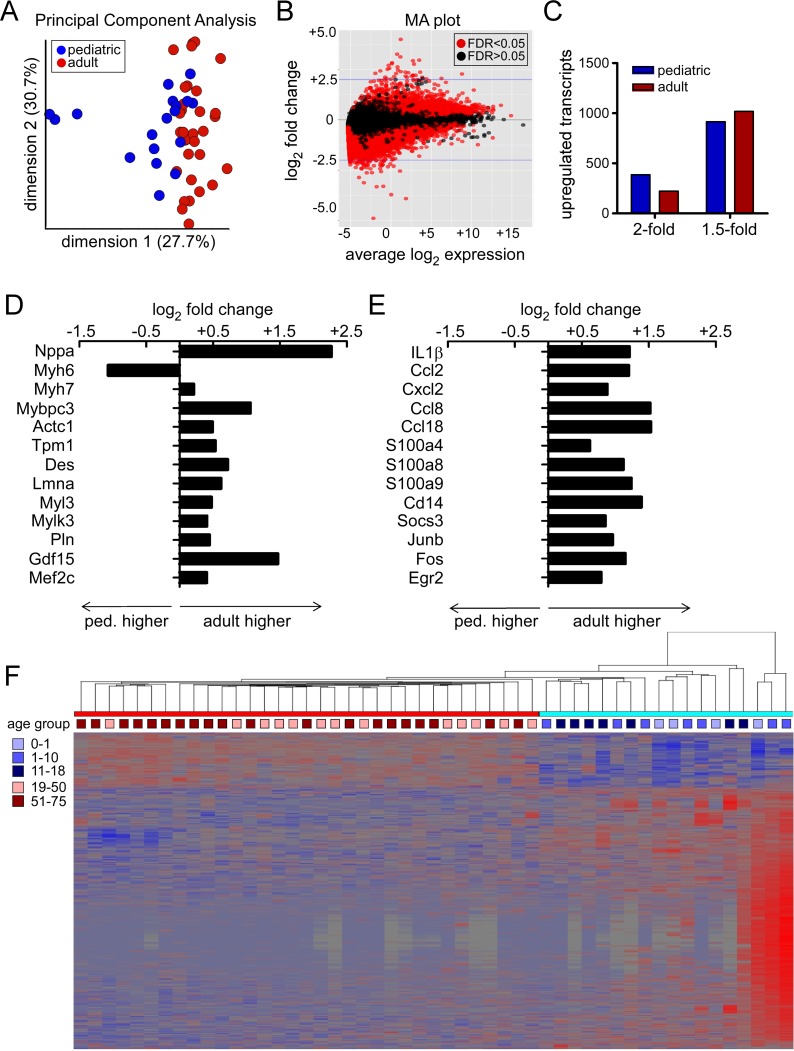
Pediatric dilated cardiomyopathy (DCM) is the most common indication for heart transplantation in children. Despite similar genetic etiologies, medications routinely used in adult heart failure patients do not improve outcomes in the pediatric population. The mechanistic basis for these observations is unknown. We hypothesized that pediatric and adult DCM comprise distinct pathological entities, in that children do not undergo adverse remodeling, the target of adult heart failure therapies. To test this hypothesis, we examined LV specimens obtained from pediatric and adult donor controls and DCM patients. Consistent with the established pathophysiology of adult heart failure, adults with DCM displayed marked cardiomyocyte hypertrophy and myocardial fibrosis compared with donor controls. In contrast, pediatric DCM specimens demonstrated minimal cardiomyocyte hypertrophy and myocardial fibrosis compared with both age-matched controls and adults with DCM. Strikingly, RNA sequencing uncovered divergent gene expression profiles in pediatric and adult patients, including enrichment of transcripts associated with adverse remodeling and innate immune activation in adult DCM specimens. Collectively, these findings reveal that pediatric and adult DCM represent distinct pathological entities, provide a mechanistic basis to explain why children fail to respond to adult heart failure therapies, and suggest the need to develop new approaches for pediatric DCM.

Keywords: Cardiology.


Conflict of interest statement

Conflict of interest: The authors have declared that no conflict of interest exists.

Figures

Figure 1
Figure 1. Absence of cardiomyocyte hypertrophy in pediatric DCM.
(A) Wheat germ agglutinin (WGA) staining (red) demonstrating that pediatric donor controls and pediatric DCM specimens display similar cardiomyocyte cross-sectional area. In contrast, adult DCM patients display increased cardiomyocyte cross-sectional area compared with donor controls. Blue, DAPI (nuclei). Magnification 400×. (B) Quantification of cardiomyocyte cross-sectional area. (C) Cardiomyocyte cross-sectional area stratified by age, revealing that cardiomyocyte cross-sectional area does not differ from donor controls across pediatric groups. *P < 0.05 (Mann Whitney U test). (D) Scatter plots showing that cardiomyocyte cross-sectional area does not differ as a function of age in pediatric DCM patients. In contrast, adult DCM patients have increased cardiomyocyte cross-sectional area at all ages examined.
Figure 2
Figure 2. Sarcomere structure in pediatric and adult DCM.
(A) Electron microscopy examining sarcomere structure in donor control, pediatric, and adult DCM specimens. Compared with donor controls, pediatric DCM patients display no change in sarcomere thickness. In contrast, adult DCM patients demonstrate increased sarcomere thickness compared with donor controls. (B) Quantification of sarcomere thickness. Each data point represents an average of >20 sarcomeres measured from individual patient samples. P values found with Mann Whitney U test.
Figure 3
Figure 3. Absence of myocardial fibrosis in pediatric DCM.
(A) Picrosirius red staining (red) demonstrating that, compared with donor controls, pediatric DCM specimens display minimal interstitial or perivascular fibrosis. In contrast, adult DCM patients display increased interstitial and perivascular fibrosis compared with donor controls. Magnification 200×. (B) Quantification of percent fibrosis as assessed by total Picrosirius red staining per 20× field in pediatric and adult control and DCM specimens. (C) Quantification of interstitial and perivascular fibrosis scores. (D) Scatter plots showing that the percent of Picrosirius staining in pediatric DCM specimens does not differ from donor pediatric controls across the spectrum of child age. In contrast, adult DCM patients have increased Picrosirius red staining at all ages examined. (E and F) Total Picrosirius red staining (E) and fibrosis scores (F) stratified by age, revealing that interstitial and perivascular fibrosis scores are only increased in adult groups. *P < 0.05 (χ2 test), ***P < 0.05 (compared to pediatric groups).
Figure 4
Figure 4. Microvascular alterations in pediatric and adult DCM.
(A) CD34 immunostaining demonstrating that, compared with donor controls, pediatric DCM specimens display increased microvascular density. In contrast, adult DCM patients have indistinguishable microvascular density compared with donor controls. Magnification 200×. (B) Quantification of microvascular density expressed as the percent of staining per 20× field. (C) Scatter plots showing that pediatric DCM patients display increased capillary density compared with donor controls across the spectrum of child ages. In contrast, capillary density did not differ between adult DCM specimens and donor controls at all ages examined. (D) CD34 staining stratified by age, revealing that microvascular density is increased in all pediatric groups. *P < 0.05 (Mann Whitney U test).
Figure 5
Figure 5. Disease duration does not influence pathological evidence of adverse remodeling in adult and pediatric DCM.
(A) Quantification of cardiomyocyte cross-sectional area stratified by disease duration reveals that patients diagnosed with heart failure < 1 year, 1–5 years, or < 5 years prior to tissue procurement displayed marked cardiomyocyte hypertrophy compared with donor controls. No significant differences were evident between disease duration groups. (B) Linear regression analysis describing the relationship between cardiomyocyte cross-sectional area and disease duration. (C) Quantification of total Picrosirius red staining stratified by disease duration reveals that patients diagnosed with heart failure < 1 year, 1–5 years, or < 5 years prior to tissue procurement displayed increased fibrosis compared with donor controls. (D) Linear regression analysis describing the relationship between total Picrosirius red staining and disease duration. (E and F) Quantification of interstitial and perivascular fibrosis stratified by disease duration demonstrates that myocardial fibrosis occurs independent of disease duration. No significant differences were evident between disease duration groups. (G and H) Quantification of CD34 staining stratified by disease duration demonstrates no relationship between microvascular density and disease duration. (I and J) Quantification of myocardial fibrosis (I) and cardiomyocyte cross-sectional area (J) stratified by disease duration reveals that children diagnosed with heart failure < 1 year or < 1 year prior to tissue procurement displayed no indistinguishable levels of myocardial fibrosis and cardiomyocyte hypertrophy compared with donor controls. No significant differences were evident between disease duration groups. *P < 0.05 compared with donor control. Mann Whitney U test (A, C, G) or χ2 test (E and F).
Figure 6
Figure 6. RNA sequencing analysis.
(A) Principal component analysis demonstrating segregation of pediatric and adult DCM samples. (B) Bland-Altman (MA) plot depicting the relationship between differential gene expression and transcript abundance in pediatric and adult DCM samples. (C) Differential gene expression analysis highlighting the number of mRNA transcripts that are differentially expressed by 1.5- and 2-fold in pediatric and adult DCM samples. (D and E) mRNA transcripts associated with sarcomere remodeling (D) and inflammation (E) are differentially regulated in pediatric and adult DCM. (F) Hierarchical clustering analysis illustrating that pediatric and adult DCM samples cluster as two distinct entities rather than as a continuum of age. Ped, pediatric.

References

    1. Towbin JA, et al. Incidence, causes, and outcomes of dilated cardiomyopathy in children. JAMA. 2006;296(15):1867–1876. doi: 10.1001/jama.296.15.1867. - DOI - PubMed
    1. Taylor DO, et al. Registry of the International Society for Heart and Lung Transplantation: Twenty-sixth Official Adult Heart Transplant Report-2009. J Heart Lung Transplant. 2009;28(10):1007–1022. doi: 10.1016/j.healun.2009.08.014. - DOI - PubMed
    1. Go AS, et al. Heart disease and stroke statistics--2013 update: a report from the American Heart Association. Circulation. 2013;127(1):e6–e245. doi: 10.1161/CIR.0b013e31828124ad. - DOI - PMC - PubMed
    1. Rampersaud E, Siegfried JD, Norton N, Li D, Martin E, Hershberger RE. Rare variant mutations identified in pediatric patients with dilated cardiomyopathy. Prog Pediatr Cardiol. 2011;31(1):39–47. doi: 10.1016/j.ppedcard.2010.11.008. - DOI - PMC - PubMed
    1. Goldberg LR. In the clinic. Heart failure. Ann Intern Med. 2010;152(11):ITC61–15; quiz ITC616. - PubMed