Review
. 2017 Jul 20;2(14):e93751.
doi: 10.1172/jci.insight.93751.

Diabetic retinopathy: current understanding, mechanisms, and treatment strategies

Affiliations
Review

Diabetic retinopathy: current understanding, mechanisms, and treatment strategies

Elia J Duh et al. JCI Insight. .

Abstract

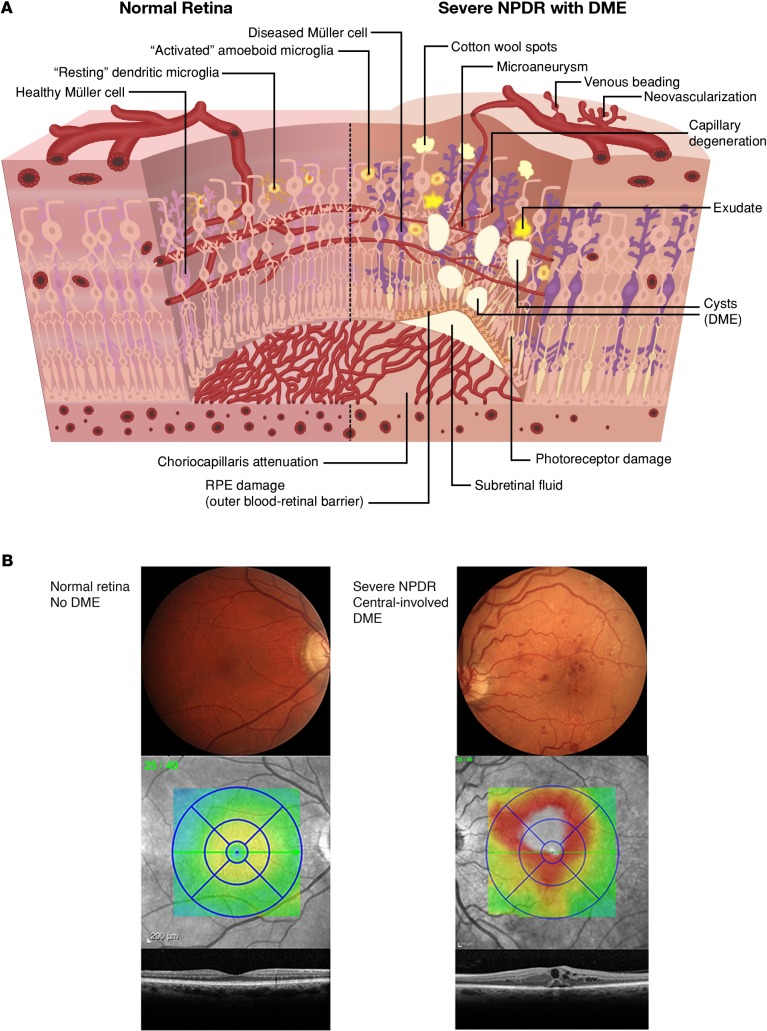
Diabetic retinopathy (DR) causes significant visual loss on a global scale. Treatments for the vision-threatening complications of diabetic macular edema (DME) and proliferative diabetic retinopathy (PDR) have greatly improved over the past decade. However, additional therapeutic options are needed that take into account pathology associated with vascular, glial, and neuronal components of the diabetic retina. Recent work indicates that diabetes markedly impacts the retinal neurovascular unit and its interdependent vascular, neuronal, glial, and immune cells. This knowledge is leading to identification of new targets and therapeutic strategies for preventing or reversing retinal neuronal dysfunction, vascular leakage, ischemia, and pathologic angiogenesis. These advances, together with approaches embracing the potential of preventative or regenerative medicine, could provide the means to better manage DR, including treatment at earlier stages and more precise tailoring of treatments based on individual patient variations.


Conflict of interest statement

Conflict of interest: The authors declare that no conflict of interest exists.

Figures

Figure 1
Figure 1. Pathological lesions of diabetic retinopathy.
(A) An illustrated schematic of normal retina compared with nonproliferative diabetic retinopathy (NPDR) with diabetic macular edema (DME). The normal healthy retina includes healthy retinal blood vessels, glial elements including Müller cells, neuronal elements including photoreceptors, and resting microglia. The inner and outer blood-retinal barriers are intact. In contrast, the retina in diabetic retinopathy exhibits multiple abnormalities, including vascular changes (microaneurysms, venous beading, capillary degeneration, and neovascularization), lesions associated with vascular damage (cotton wool spots and exudate), glial dysfunction including Müller cell swelling, neuronal damage, activated microglia, retinal pigment epithelium (RPE) damage, and thinning of the choriocapillaris. There is dysfunction of the inner and outer blood-retinal barrier, with resulting accumulation of fluid in the retina, which can be manifested by thickening of retinal layers, cysts, and subretinal fluid. (B) Color fundus photograph and OCT images of a normal healthy retina and retina with severe NPDR with central-involved DME are shown. Illustrated by Rachel Davidowitz.
Figure 2
Figure 2. The neurovascular unit and its disruption by diabetes.
In normal, healthy retina (shown in the center), there is functional coupling and interdependency of neurons, glial elements including Müller cells, and vascular cells, with associated immune cells such as microglia. The insets show pathological changes associated with diabetic retinopathy in multiple components of the neurovascular unit and interacting immune cells, including compromise of endothelial-mural cell interactions, vascular basement membrane damage, Müller cell gliosis, and immune cell activation. Together, these changes result in impairment of neurovascular coupling, with consequences including blood-retinal barrier breakdown and dysregulation of retinal blood flow. Illustrated by Rachel Davidowitz.

References

    1. Guariguata L, Whiting DR, Hambleton I, Beagley J, Linnenkamp U, Shaw JE. Global estimates of diabetes prevalence for 2013 and projections for 2035. Diabetes Res Clin Pract. 2014;103(2):137–149. doi: 10.1016/j.diabres.2013.11.002. - DOI - PubMed
    1. Nanditha A, et al. Diabetes in Asia and the Pacific: Implications for the Global Epidemic. Diabetes Care. 2016;39(3):472–485. doi: 10.2337/dc15-1536. - DOI - PubMed
    1. Stitt AW, et al. The progress in understanding and treatment of diabetic retinopathy. Prog Retin Eye Res. 2016;51:156–186. doi: 10.1016/j.preteyeres.2015.08.001. - DOI - PubMed
    1. Antonetti DA, Klein R, Gardner TW. Diabetic retinopathy. N Engl J Med. 2012;366(13):1227–1239. doi: 10.1056/NEJMra1005073. - DOI - PubMed
    1. Klein R, Klein BE. Are individuals with diabetes seeing better?: a long-term epidemiological perspective. Diabetes. 2010;59(8):1853–1860. doi: 10.2337/db09-1904. - DOI - PMC - PubMed