. 2017 Jul 19;7(1):5862.
doi: 10.1038/s41598-017-06180-5.

Generally Applicable Transformation Protocols for Fluorescent Nanodiamond Internalization into Cells

Affiliations

Generally Applicable Transformation Protocols for Fluorescent Nanodiamond Internalization into Cells

Simon R Hemelaar et al. Sci Rep. .

Abstract

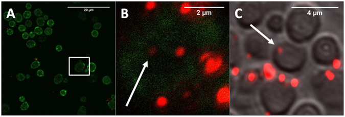
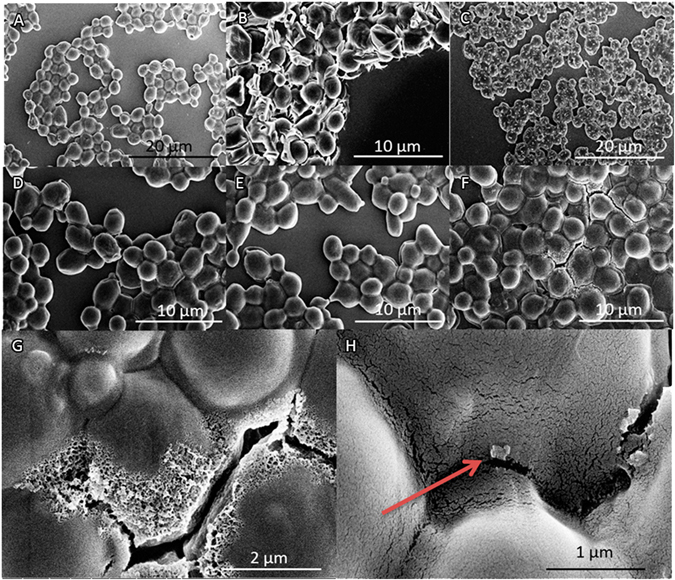
Fluorescent nanodiamonds (FNDs) are promising nanoprobes, owing to their stable and magnetosensitive fluorescence. Therefore they can probe properties as magnetic resonances, pressure, temperature or strain. The unprecedented sensitivity of diamond defects can detect the faint magnetic resonance of a single electron or even a few nuclear spins. However, these sensitivities are only achieved if the diamond probe is close to the molecules that need to be detected. In order to utilize its full potential for biological applications, the diamond particle has to enter the cell. Some model systems, like HeLa cells, readily ingest particles. However, most cells do not show this behavior. In this article we show for the first time generally applicable methods, which are able to transport fluorescent nanodiamonds into cells with a thick cell wall. Yeast cells, in particular Saccharomyces cerevisiae, are a favored model organism to study intracellular processes including aging on a cellular level. In order to introduce FNDs in these cells, we evaluated electrical transformation and conditions of chemical permeabilization for uptake efficiency and viability. 5% DMSO (dimethyl sulfoxide) in combination with optimized chemical transformation mix leads to high uptake efficiency in combination with low impact on cell biology. We have evaluated all steps in the procedure.


Conflict of interest statement

The authors declare that they have no competing interests.

Figures

Figure 1
Figure 1
Qualitative analysis of uptake of FNDs by Hxt6-GFP-expressing yeast cells. (A) Overview of FNDs and Hxt6-GFP-expressing cells after treatment with TMIX, incubation at 42 °C and treatment with 5% DMSO. (B) Close up of the boxed cell from A, with diamond particles inside (for better visibility of the diamond particles the contrast of the red channel has been increased). The arrow shows a particle that was not at any place associated with the cell membrane. (C) Embedded and sectioned (approximately 0.8 µm thickness) cells, visualized using Differential Interference Contrast. The diamond particle indicated by the arrow is internalized.
Figure 2
Figure 2
Quantitative analysis of FND uptake by Hxt6-GFP-expressing Saccharomyces cerevisiae cells. The amount of objects (adjacent FND positive pixels are counted as an object) and particles (an object can also be an aggregate consisting of more than one particle) is estimated through our home written FIJI protocol (see Method section: FND uptake quantification). For all situations, 2 times approximately 100 cells were selected and cells with obvious large aggregates on the exterior were excluded post-hoc. (A and B) show the absolute numbers for both types of transformations. In (C and D) a grouped distribution of the percentage of cells carrying a range of objects is shown. Significance is tested compared to the control situation. *p < 0.05, ***p < 0.001, ****p < 0.0001.
Figure 3
Figure 3
Survival of yeast cells after performing the interventions and different steps in the transformation protocols separately. Colony forming units (CFUs) are a measure for the survival of viable cells after either the chemical transformation or the electroporation protocol. (A) The greatest reduction of viability occurs after the addition of 0.01% Triton. The addition of FNDs to the treatment does not decrease the viability. Bars represent averages of triplicates out of two independent experiments. An area of +/−20% around 100% is deemed as ‘normal viability,’ (B). Electroporation reduces viability by a factor 102. In all samples FNDs were also added. In the case of 8 pulses, cell viability was completely reduced. For the electroporation protocol all decreases were significant (p < 0.0001). Bars represent averages of replicates out of two independent experiments, error bars show the Standard Error of the Mean. Significance is tested compared to the control situation. *p < 0.05, ***p < 0.001.
Figure 4
Figure 4
SEM visualizations of yeast cell topography and morphology. (A) Cells incubated with FNDs. (B) Cells after treatment with 2% DMSO and TMIX. Distorted cell morphology shows the side effects of this technique. (C) Cells treated with 0.1% Triton and FNDs: an accumulation of salts due to drying on the outside of the cells can be seen in white. (D) Cells treated with 0.1% Triton, TMIX and FNDs. (E) Cells electroporated with 1 pulse. Some light cellular damage can be observed. (F) Cells electroporated with 8 pulses. The cell wall of these cells is severely damaged as a result of the applied high electrical currents. (G) Disintegration of the cell wall results in puncturing and will lead to cell death (close up of F). (H) Close up of a diamond particle on the outside of a cell (indicated by the arrow, close up of A).

References

    1. Mohan N, Chen CS, Hsieh HH, Wu YC, Chang HC. In vivo imaging and toxicity assessments of fluorescent nanodiamonds in caenorhabditis elegans. Nano Lett. 2010;10:3692–3699. doi: 10.1021/nl1021909. - DOI - PubMed
    1. Schirhagl R, Chang K, Loretz M, Degen CL. Nitrogen-vacancy centers in diamond: nanoscale sensors for physics and biology. Annu. Rev. Phys. Chem. 2014;65:83–105. doi: 10.1146/annurev-physchem-040513-103659. - DOI - PubMed
    1. Neumann P, et al. High-precision nanoscale temperature sensing using single defects in diamond. Nano Lett. 2013;13:2738–2742. doi: 10.1021/nl401216y. - DOI - PubMed
    1. Grinolds MS, et al. Nanoscale magnetic imaging of a single electron spin under ambient conditions. Nat. Phys. 2013;9:215–219. doi: 10.1038/nphys2543. - DOI
    1. Hemelaar, S. R. et al. The interaction of fluorescent nanodiamond probes with cellular media. Microchim. Acta 1–9 (2017). doi:10.1007/s00604-017-2086-6 - PMC - PubMed

LinkOut - more resources