. 2017 Jul 19;7(1):5887.
doi: 10.1038/s41598-017-06178-z.

Endothelial Dysfunction in Severe Preeclampsia is Mediated by Soluble Factors, Rather than Extracellular Vesicles

Affiliations

Endothelial Dysfunction in Severe Preeclampsia is Mediated by Soluble Factors, Rather than Extracellular Vesicles

Michelle O'Brien et al. Sci Rep. .

Abstract

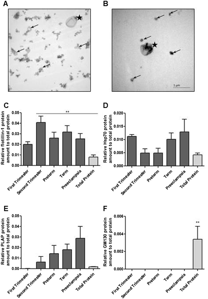
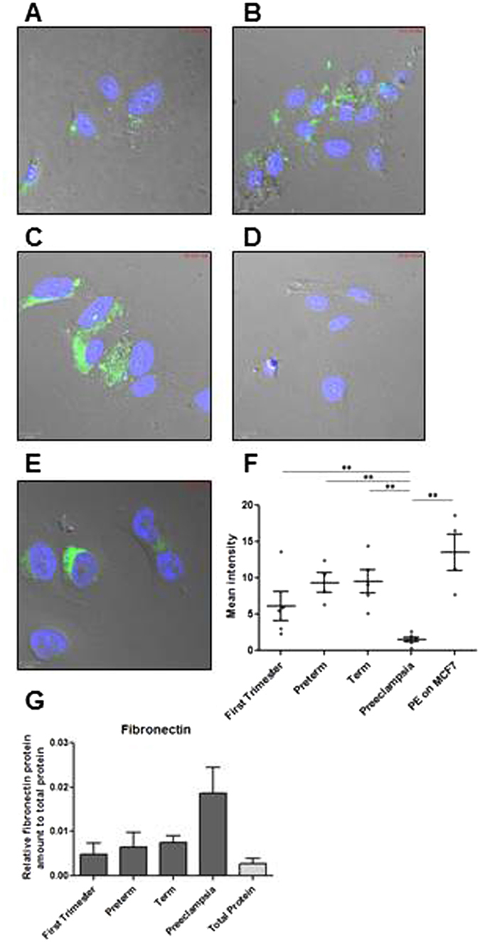
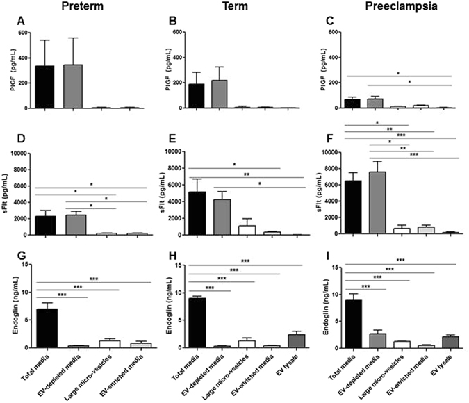
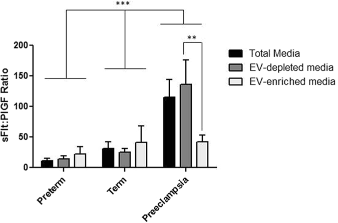
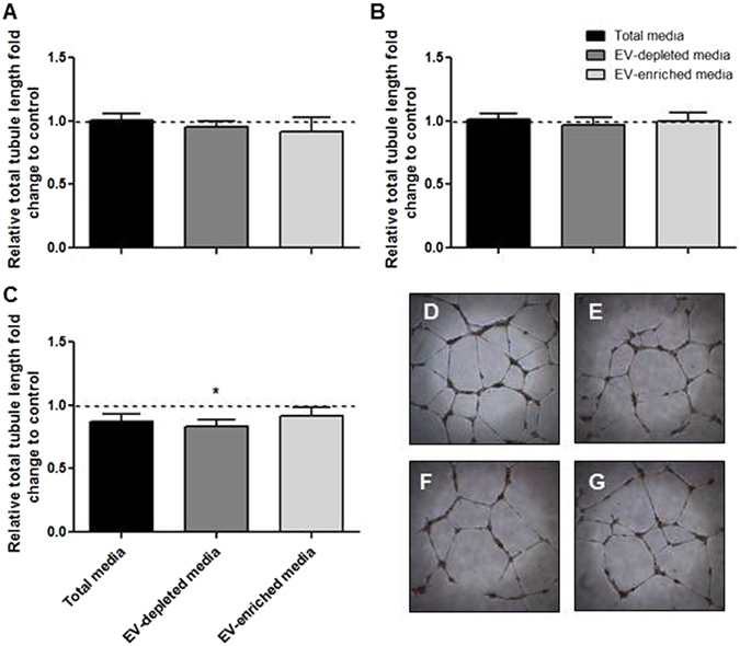
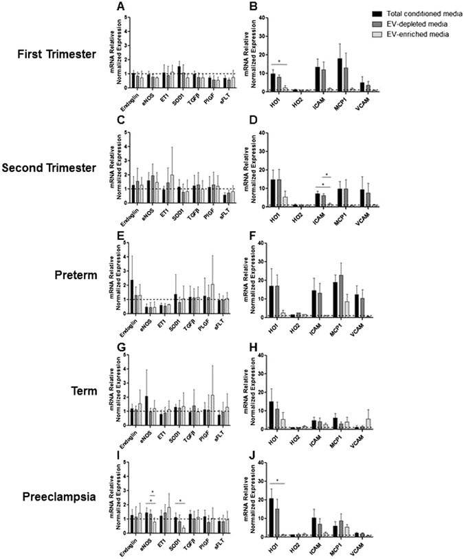
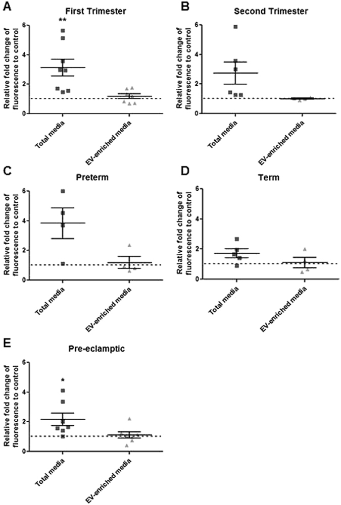
In severe early-onset preeclampsia (sPE) the placenta releases soluble angiogenesis-regulating proteins, trophoblast-derived fragments, and extracellular vesicles (EVs). Their relative importance in disease pathogenesis is not presently understood. We explanted placental villi from healthy and sPE women then separated the media into: total-conditioned, EV-depleted and EV-enriched media. Three fractions were compared for; angiogenic protein secretion by ELISA, angiogenic and inflammation gene mRNA expression and leukocyte adhesion assay. sPE placental villi secreted significantly less PlGF (70 ± 18 pg/mL) than preterm controls (338 ± 203; p = 0.03). sFlt-1:PlGF ratios in total-conditioned (115 ± 29) and EV-depleted media (136 ± 40) from sPE placental villi were significantly higher than in EV-enriched media (42 ± 12; p < 0.01) or any preterm or term media. Fluorescent-labeled EVs derived across normal gestation, but not from sPE, actively entered HUVECs. From sPE placental villi, the soluble fraction, but not EV-enriched fraction, significantly repressed angiogenesis (0.83 ± 0.05 fold, p = 0.02), induced HO-1 mRNA (15.3 ± 5.1 fold, p < 0.05) and induced leukocyte adhesion (2.2 ± 0.4 fold, p = 0.04). Soluble media (total-conditioned and EV-depleted media) from sPE placental villi induced endothelial dysfunction in HUVEC, while the corresponding EV-enriched fraction showed no such effects. Our data suggest that soluble factors including angiogenesis-regulating proteins, dominate the vascular pathology of this disease.


Conflict of interest statement

The authors declare that they have no competing interests.

Figures

Figure 1
Figure 1
Purity of EVs was assessed using low magnification TEM. (A) EV preparation prior to and (B) following qEV exclusion column. Images taken at 19,000X magnification. Arrows = exosome, * = micro-vesicles. (CF) Additional characterization of the EVs was performed by Western blotting for EV-specific markers, Flotilin-1 and Hsp70, and for placenta specific marker-PLAP. Purity of preparations was further validated with GM130 antibody. Densitometry analysis for (C) Flotilin-1, (D) Hsp70, (E) PLAP and (F) GM130 are presented.
Figure 2
Figure 2
PKH67 fluorescently labelled EVs generated from (A) first, (B) preterm, (C) term and (D) PE placental explants were incubated with HUVEC cells for 4 hrs. (E) Pre-eclamptic EVs were also incubated with MCF7 breast cancer cell line for 4 hrs. (F) PE-derived EVs are unable to enter HUVEC cells but are able to enter into cancer cells. (G) EVs generated from PE express fibronectin.
Figure 3
Figure 3
Media analysis by ELISA for (AC) PlGF, (DF) sFlt-1 and (GI) Endoglin from preterm, term and PE placenta. PlGF and sFlt-1 levels were mainly detected in total and EV-free media. Endoglin was detected in total media but its levels were markedly reduced in EV-depleted media. All three angiogenic proteins showed minimal levels in EV-enriched media, microvesicle-enriched media and EV lysates. Large micro-vesicles = obtained after the 10,000 g spin, include micro-vesicle and apoptotic bodies. EV lysates = obtained by RIPA lysis of purified EV pellets.
Figure 4
Figure 4
sFLt-1:PlGF ratio in media preparations from preterm, term and sPE placentas. NS = not significant.
Figure 5
Figure 5
Angiogenic activity of HUVECs treated with (A) preterm or (B) term conditioned and enriched media was not altered. (C) HUVEC angiogenesis was moderately decreased when treated with PE conditioned media but not EV-enriched media. Representative images of (D) positive control, (E) total PE media, (F) EV-depleted media and (G) PE-EV enriched media.
Figure 6
Figure 6
HUVECs were treated with total, EV-depleted and EV-enriched media from (A,B) first trimester, (C,D) second trimester, (E,F) preterm, (G,H) term and (I,J) PE placentas for 24 hr.
Figure 7
Figure 7
Endothelial cells were treated with total and EV-enriched media from (A) first trimester, (B) second trimester, (C) preterm, (D) term and (E) PE placentas for 6 hrs prior to incubation with fluorescently labelled monocytes for 45 min. First trimester and PE total media but not the EV-enriched media elicited leukocyte activation.

References

    1. Mol BW, et al. Pre-eclampsia. Lancet. 2016;387:999–1011. doi: 10.1016/S0140-6736(15)00070-7. - DOI - PubMed
    1. Karumanchi SA, Granger JP. Preeclampsia and Pregnancy-Related Hypertensive Disorders. Hypertension. 2016;67:238–242. doi: 10.1161/HYPERTENSIONAHA.116.06421. - DOI - PMC - PubMed
    1. Walker MG, et al. Sex-specific basis of severe placental dysfunction leading to extreme preterm delivery. Placenta. 2012;33:568–571. doi: 10.1016/j.placenta.2012.03.011. - DOI - PubMed
    1. Redline RW. Classification of placental lesions. American journal of obstetrics and gynecology. 2015;213:S21–28. doi: 10.1016/j.ajog.2015.05.056. - DOI - PubMed
    1. Rajakumar A, et al. Transcriptionally active syncytial aggregates in the maternal circulation may contribute to circulating soluble fms-like tyrosine kinase 1 in preeclampsia. Hypertension. 2012;59:256–264. doi: 10.1161/HYPERTENSIONAHA.111.182170. - DOI - PMC - PubMed

Publication types

Substances

Grants and funding

LinkOut - more resources