Review
. 2017 Jun;6(3):499-509.
doi: 10.21037/tau.2017.05.31.

Is contrast enhancement needed for diagnostic prostate MRI?

Affiliations
Review

Is contrast enhancement needed for diagnostic prostate MRI?

Michele Scialpi et al. Transl Androl Urol. 2017 Jun.

Abstract

Prostate Imaging Reporting and Data System version 2 (PI-RADS v2) provides clinical guidelines for multiparametric magnetic resonance imaging (mpMRI) [T2-weighted imaging (T2WI), diffusion-weighted imaging (DWI) and dynamic contrast-enhanced magnetic resonance imaging (DCE-MRI)] of prostate. However, DCE-MRI seems to show a limited contribution in prostate cancer (PCa) detection and management. In our experience, DCE-MRI, did not show significant change in diagnostic performance in addition to DWI and T2WI [biparametric MRI (bpMRI)] which represent the predominant sequences to detect suspected lesions in peripheral and transitional zone (TZ). In this article we reviewed the role of DCE-MRI also indicating the potential contribute of bpMRI approach (T2WI and DWI) and lesion volume evaluation in the diagnosis and management of suspected PCa.

Keywords: Prostate Imaging Reporting and Data System version 2 (PI-RADS v2); Prostate cancer (PCa); biparametric magnetic resonance imaging (bpMRI).


Conflict of interest statement

Conflicts of Interest: The authors have no conflicts of interest to declare.

Figures

Figure 1
Figure 1
A 72-year-old patient with PSA of 7.3 ng/mL but without previous prostate biopsy. Focal lesion of 10 mm (arrow) in the base in the right side of the TZ with moderately low signal intensity on T2WI (A), high signal intensity on high b-value image of the DWI (B), low signal on ADC map (C) and moderately contrast enhancement on DCE (D). The lesion was assigned a PI-RADS v2 score of 3. The enhancement on DCE was considered irrelevant and biopsy was required because of the suspicious lesion. Targeted biopsy revealed a Gleason 3+3 prostate cancer. PSA, prostate-specific antigen; TZ, transitional zone; T2WI, T2-weighted imaging; DWI, diffusion-weighted imaging; ADC, apparent diffusion coefficient; DCE, dynamic contrast-enhanced, PI-RADS, prostate imaging reporting and data system.
Figure 2
Figure 2
Variability of index tumor and index lesion diameter measured by histology and MRI respectively for GS =6 and GS ≥7 groups. In horizontal graph lines, boxes and whiskers represent the median, interquartile range, and range values respectively. Comparison between GS =6 and GS >7 was performed using the nonparametric Mann-Whitney test. **, P=0.008. MRI, magnetic resonance imaging.
Figure 3
Figure 3
The area under the ROC curve (performed by comparing values of tumor lesion volume in GS 7 and GS =6 groups) was 0.752 (P<0.0001) and lesion volume showed a sensitivity and specificity of 80% and 67% respectively in significant PCa prediction.
Figure 4
Figure 4
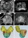
A 67-year-old patient with PSA of 4.1 ng/mL but without previous prostate biopsy. Focal lesion of 12 mm (arrow) in the anterior TZ of the base of the prostate in the right side with moderately low signal intensity on T2WI (A), high signal intensity on high B value image of the DWI (B), low signal on ADC map (C) and moderately contrast enhancement on DCE (D). 3D reconstructions (E,F) showed a lesion volume <0.5 mL. The lesion was assigned a PI-RADS v2 score of 3. The enhancement on DCE was considered irrelevant and biopsy was required because of the suspicious lesion. Targeted biopsy revealed fibrous hyperplasia. PSA, prostate-specific antigen; TZ, transitional zone; T2WI, T2-weighted imaging; DWI, diffusion-weighted imaging; ADC, apparent diffusion coefficient; DCE, dynamic contrast-enhanced; PI-RADS, prostate imaging reporting and data system.
Figure 5
Figure 5
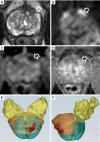
A 59-year-old patient with PSA of 7.6 ng/mL but without previous prostate biopsy. Focal lesion of 12 mm (arrow) in the anterior and posterior TZ in the base of the prostate on the right side with moderately low signal intensity on T2WI (A), high signal intensity on high B value image of the DWI (B), low signal on ADC map (C) and moderately contrast enhancement on DCE (D). 3D reconstructions (E,F) showed a lesion volume >0.5 mL. The lesion was assigned a PI-RADS v2 score of 4. The enhancement on DCE was considered irrelevant and on the basis of lesion volume biopsy was required. Targeted biopsy revealed a Gleason 3+3 prostate cancer. PSA, prostate-specific antigen; TZ, transitional zone; T2WI, T2-weighted imaging; DWI, diffusion-weighted imaging; ADC, apparent diffusion coefficient; DCE, dynamic contrast-enhanced; PI-RADS v2, prostate imaging reporting and data system version 2.
Figure 6
Figure 6
A 60-year-old patient with PSA of 6.8 ng/mL but without previous prostate biopsy. Focal lesion of 15 mm (arrow) in the anterior fibromuscular stroma of the mid-prostate on the left side with moderately low signal intensity on T2WI (A), high signal intensity on high B value image of the DWI (B), low signal on ADC map (C) and moderately contrast enhancement on DCE (D). 3D reconstructions (E,F) showed a lesion volume >0.5 mL. The lesion was assigned a PI-RADS v2 score of 4. The enhancement on DCE was considered irrelevant and on the basis of lesion volume biopsy was required. Targeted biopsy revealed a Gleason 3+4 prostate cancer. PSA, prostate-specific antigen; TZ, transitional zone; T2WI, T2-weighted imaging; DWI, diffusion-weighted imaging; ADC, apparent diffusion coefficient; DCE, dynamic contrast-enhanced; PI-RADS v2, prostate imaging reporting and data system version 2.

References

    1. Prostate Imaging Reporting and Data System (PI-RADS). Reston (VA): American College of Radiology. Available online: http://www.acr.org/Quality-Safety/ Resources/PIRADS/
    1. Rosenkrantz AB, Oto A, Turkbey B, et al. Prostate Imaging Reporting and Data System (PI-RADS), Version 2: A Critical Look. AJR Am J Roentgenol 2016;206:1179-83. 10.2214/AJR.15.15765 - DOI - PubMed
    1. Abd-Alazeez M, Kirkham A, Ahmed HU, et al. Performance of multiparametric MRI in men at risk of prostate cancer before the first biopsy: a paired validating cohort study using template prostate mapping biopsies as the reference standard. Prostate Cancer Prostatic Dis 2014;17:40-6. 10.1038/pcan.2013.43 - DOI - PMC - PubMed
    1. Renard-Penna R, Roupret M, Compérat E, et al. Relationship between non-suspicious MRI and insignificant prostate cancer: results from a monocentric study. World J Urol 2016;34:673-8. 10.1007/s00345-015-1685-2 - DOI - PubMed
    1. Scialpi M, Falcone G, Scialpi P, et al. Biparametric MRI: a further improvement to PIRADS 2.0? Diagn Interv Radiol 2016;22:297-8. 10.5152/dir.2016.15598 - DOI - PMC - PubMed