Review
. 2017 Jun 29:4:88.
doi: 10.3389/fmed.2017.00088. eCollection 2017.

Imaging Bronchopulmonary Dysplasia-A Multimodality Update

Affiliations
Review

Imaging Bronchopulmonary Dysplasia-A Multimodality Update

Thomas Semple et al. Front Med (Lausanne). .

Abstract

Bronchopulmonary dysplasia is the most common form of infantile chronic lung disease and results in significant health-care expenditure. The roles of chest radiography and computed tomography (CT) are well documented but numerous recent advances in imaging technology have paved the way for newer imaging techniques including structural pulmonary assessment via lung magnetic resonance imaging (MRI), functional assessment via ventilation, and perfusion MRI and quantitative imaging techniques using both CT and MRI. New applications for ultrasound have also been suggested. With the increasing array of complex technologies available, it is becoming increasingly important to have a deeper knowledge of the technological advances of the past 5-10 years and particularly the limitations of some newer techniques currently undergoing intense research. This review article aims to cover the most salient advances relevant to BPD imaging, particularly advances within CT technology, postprocessing and quantitative CT; structural MRI assessment, ventilation and perfusion imaging using gas contrast agents and Fourier decomposition techniques and lung ultrasound.

Keywords: bronchopulmonary dysplasia; hyperpolarized gas imaging; imaging techniques; lung parenchymal magnetic resonance imaging; lung ultrasound; quantitative pulmonary magnetic resonance imaging; structural characterization.


Figures

Figure 1
Figure 1
Chest radiograph demonstrating widespread coarse interstitial markings, atelectasis, and regions of hyperexpansion (particularly at the left lung base), typical of bronchopulmonary dysplasia. Note also the right upper lobe consolidation and malposition of the NG tube.
Figure 2
Figure 2
Axial computed tomography section through the upper lobes on lung window settings demonstrates linear and subpleural opacities, bronchial wall thickening, and areas of low attenuation (indicative of small airways disease) in a patient with bronchopulmonary dysplasia.
Figure 3
Figure 3
Vacuum immobilization device used to limit gross patient movement. Use of these devices, alongside ultrafast, high-pitch computed tomography, has dramatically reduced the need for general anesthetic or sedation for cardiothoracic CT at our institution.
Figure 4
Figure 4
Minimum intensity projection CT reconstruction demonstrating airway morphology and regions of heterogeneous (mosaic) attenuation in a child with bronchopulmonary dysplasia.
Figure 5
Figure 5
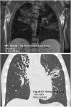
(A) Coronal black blood SSFP magnetic resonance (MR) image and (B) coronal computed tomography (CT) reconstruction in a child with cystic fibrosis. Although the spatial resolution of MRI is relatively poor compared to CT, MRI is capable of demonstrating gross airway pathology.
Figure 6
Figure 6
Magnetic resonance imaging angiogram in a child with bronchopulmonary dysplasia demonstrating poor perfusion of the right upper lobe related to severe small airways disease and reflex vasoconstriction.

Comment in

References

    1. Gortner L, Misselwitz B, Milligan D, Zeitlin J, Kollée L, Boerch K, et al. Rates of bronchopulmonary dysplasia in very preterm neonates in Europe: results from the MOSAIC cohort. Neonatology (2011) 99(2):112–7.10.1159/000313024 - DOI - PubMed
    1. Russell RB, Green NS, Steiner CA, Meikle S, Howse JL, Poschman K, et al. Cost of hospitalization for pre-term and low birth weight infants in the United States. Pediatrics (2007) 120:e1–9.10.1542/peds.2006-2386 - DOI - PubMed
    1. Barradas DT, Wasserman MP, Daniel-Robinson L, Bruce MA, DiSantis KI, Navarro FH, et al. Hospital utilization and costs among preterm infants by payer: nationwide inpatient sample, 2009. Matern Child Health J (2016) 20:808–18.10.1007/s10995-015-1911-y - DOI - PMC - PubMed
    1. Jobe AH. Bronchopulmonary dysplasia. Am J Respir Crit Care Med (2001) 163:1723–9.10.1164/ajrccm.163.7.2011060 - DOI - PubMed
    1. Cherukupalli K, Larson JE, Rotschild A, Thurlbeck WM. Biochemical, clinical and morphologic studies on lungs of infants with bronchopulmonary dysplasia. Pediatr Pulmonol (1996) 22:215–29.10.1002/(SICI)1099-0496(199610)22:4<215::AID-PPUL1>3.0.CO;2-L - DOI - PubMed

LinkOut - more resources