

Spectral performance of a whole-body research photon counting detector CT: quantitative accuracy in derived image sets
- PMID: 28726669
- PMCID: PMC5565680
- DOI: 10.1088/1361-6560/aa8103
Spectral performance of a whole-body research photon counting detector CT: quantitative accuracy in derived image sets
Abstract
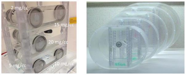
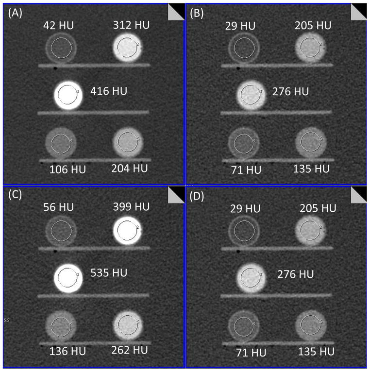
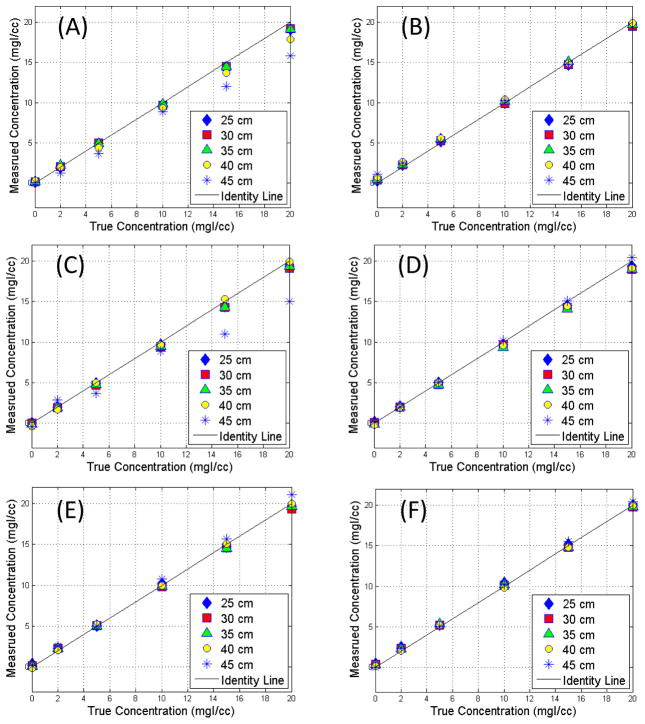
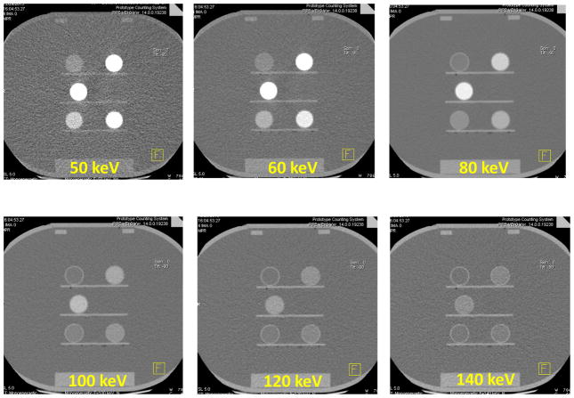
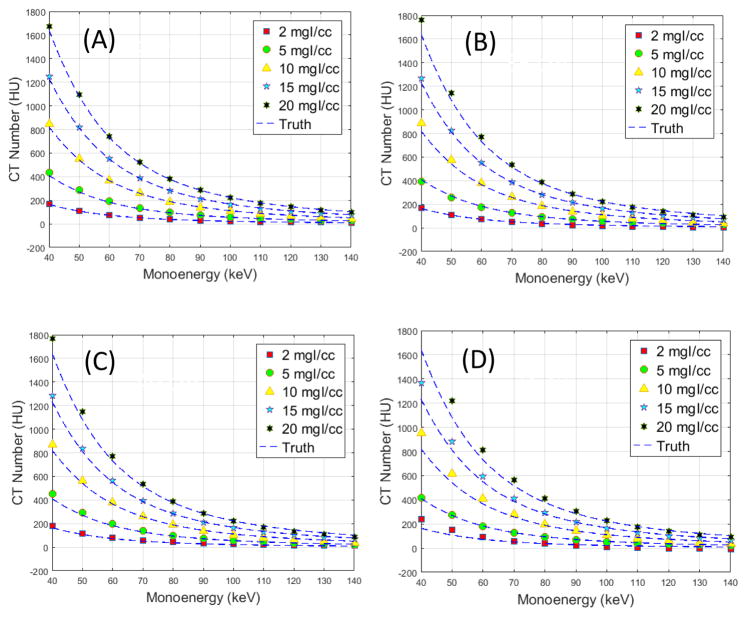
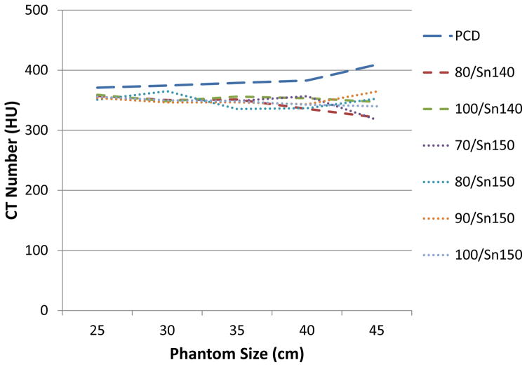
Photon-counting computed tomography (PCCT) uses a photon counting detector to count individual photons and allocate them to specific energy bins by comparing photon energy to preset thresholds. This enables simultaneous multi-energy CT with a single source and detector. Phantom studies were performed to assess the spectral performance of a research PCCT scanner by assessing the accuracy of derived images sets. Specifically, we assessed the accuracy of iodine quantification in iodine map images and of CT number accuracy in virtual monoenergetic images (VMI). Vials containing iodine with five known concentrations were scanned on the PCCT scanner after being placed in phantoms representing the attenuation of different size patients. For comparison, the same vials and phantoms were also scanned on 2nd and 3rd generation dual-source, dual-energy scanners. After material decomposition, iodine maps were generated, from which iodine concentration was measured for each vial and phantom size and compared with the known concentration. Additionally, VMIs were generated and CT number accuracy was compared to the reference standard, which was calculated based on known iodine concentration and attenuation coefficients at each keV obtained from the U.S. National Institute of Standards and Technology (NIST). Results showed accurate iodine quantification (root mean square error of 0.5 mgI/cc) and accurate CT number of VMIs (percentage error of 8.9%) using the PCCT scanner. The overall performance of the PCCT scanner, in terms of iodine quantification and VMI CT number accuracy, was comparable to that of EID-based dual-source, dual-energy scanners.
Figures


References
-
- ALMEIDA IP, SCHYNS LE, ÖLLERS MC, VAN ELMPT W, PARODI K, LANDRY G, VERHAEGEN F. Dual - energy CT quantitative imaging: a comparison study between twin - beam and dual - source CT scanners. Medical Physics 2016 - PubMed
-
- ALVAREZ RE, MACOVSKI A. Energy-selective reconstructions in x-ray computerised tomography. Physics in medicine and biology. 1976;21:733. - PubMed
-
- BALLABRIGA R, ALOZY J, CAMPBELL M, FROJDH E, HEIJNE E, KOENIG T, LLOPART X, MARCHAL J, PENNICARD D, POIKELA T. Review of hybrid pixel detector readout ASICs for spectroscopic X-ray imaging. Journal of Instrumentation. 2016;11:P01007.
-
- BENNETT JR, OPIE AM, XU Q, YU H, WALSH M, BUTLER A, BUTLER P, CAO G, MOHS A, WANG G. Hybrid spectral micro-CT: system design, implementation, and preliminary results. IEEE transactions on bio-medical engineering. 2014;61:246–53. - PubMed
-
- BOLL DT, MERKLE EM, PAULSON EK, FLEITER TR. Coronary stent patency: dual-energy multidetector CT assessment in a pilot study with anthropomorphic phantom. Radiology. 2008;247:687–95. - PubMed
MeSH terms
Substances
Grants and funding
LinkOut - more resources
Full Text Sources
Other Literature Sources
Medical