Clinical Trial
. 2017 Jul 20;8(7):e2951.
doi: 10.1038/cddis.2017.348.

Humanin G (HNG) protects age-related macular degeneration (AMD) transmitochondrial ARPE-19 cybrids from mitochondrial and cellular damage

Affiliations
Clinical Trial

Humanin G (HNG) protects age-related macular degeneration (AMD) transmitochondrial ARPE-19 cybrids from mitochondrial and cellular damage

Sonali Nashine et al. Cell Death Dis. .

Abstract

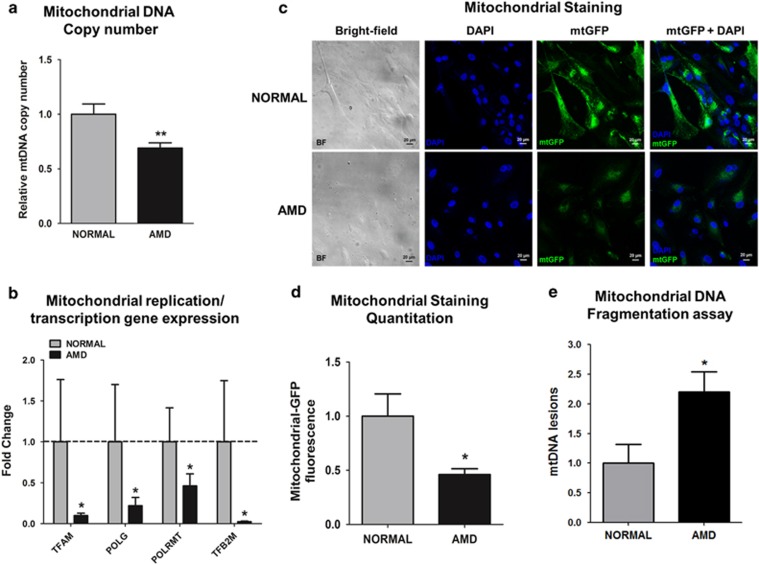
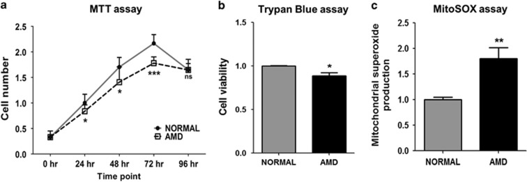
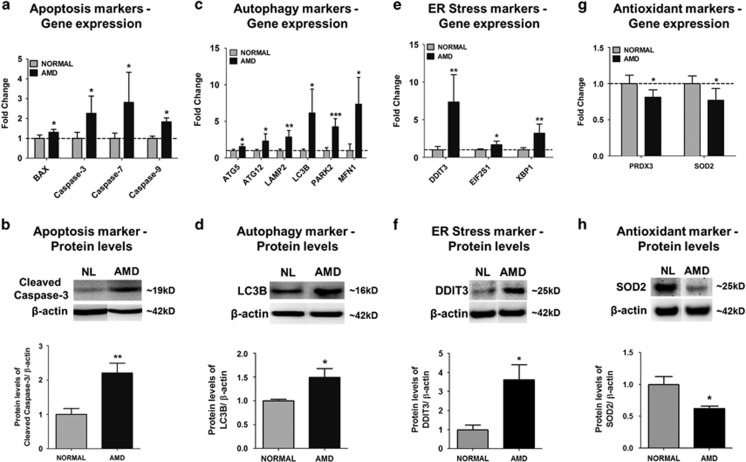
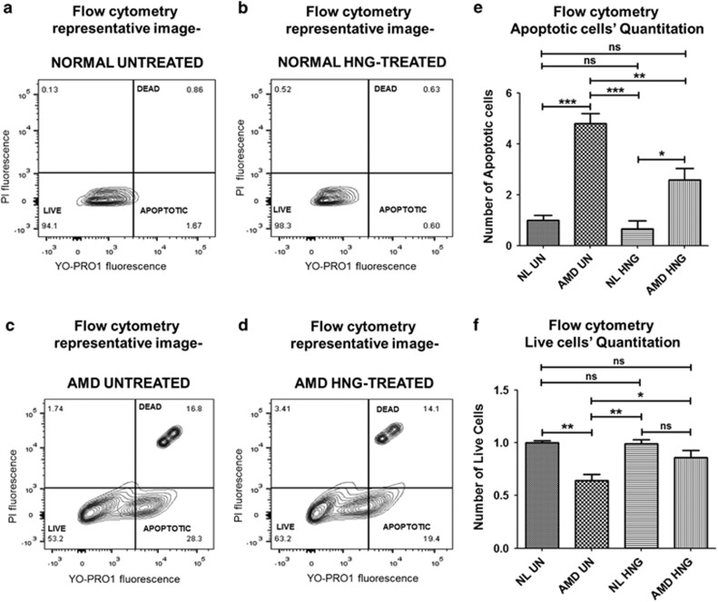
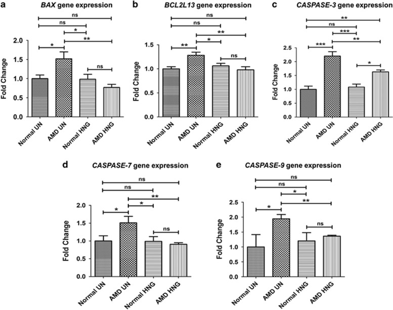
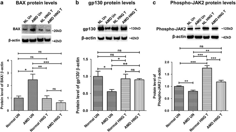
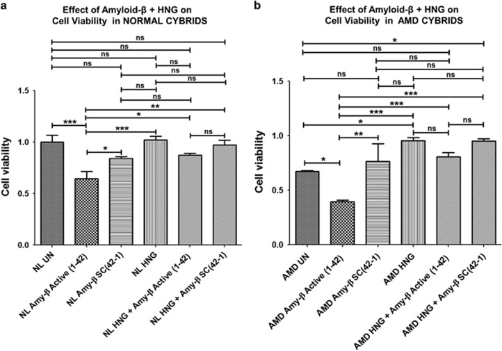
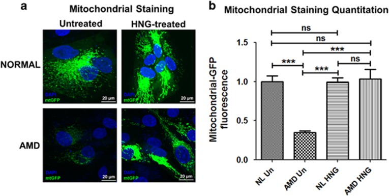
Age-related macular degeneration (AMD) ranks third among the leading causes of visual impairment with a blindness prevalence rate of 8.7%. Despite several treatment regimens, such as anti-angiogenic drugs, laser therapy, and vitamin supplementation, being available for wet AMD, to date there are no FDA-approved therapies for dry AMD. Substantial evidence implicates mitochondrial damage and retinal pigment epithelium (RPE) cell death in the pathogenesis of AMD. However, the effects of AMD mitochondria and Humanin G (HNG), a more potent variant of the mitochondrial-derived peptide (MDP) Humanin, on retinal cell survival have not been elucidated. In this study, we characterized mitochondrial and cellular damage in transmitochondrial cybrid cell lines that contain identical nuclei but possess mitochondria from either AMD or age-matched normal (Older-normal (NL)) subjects. AMD cybrids showed (1) reduced levels of cell viability, lower mtDNA copy numbers, and downregulation of mitochondrial replication/transcription genes and antioxidant enzyme genes; and (2) elevated levels of genes related to apoptosis, autophagy and ER-stress along with increased mtDNA fragmentation and higher susceptibility to amyloid-β-induced toxicity compared to NL cybrids. In AMD cybrids, HNG protected the AMD mitochondria, reduced pro-apoptosis gene and protein levels, upregulated gp130 (a component of the HN receptor complex), and increased the protection against amyloid-β-induced damage. In summary, in cybrids, damaged AMD mitochondria mediate cell death that can be reversed by HNG treatment. Our results also provide evidence of Humanin playing a pivotal role in protecting cells with AMD mitochondria. In the future, it may be possible that AMD patient's blood samples containing damaged mitochondria may be useful as biomarkers for this condition. In conclusion, HNG may be a potential therapeutic target for treatment of dry AMD, a debilitating eye disease that currently has no available treatment. Further studies are needed to establish HNG as a viable mitochondria-targeting therapy for dry AMD.


Conflict of interest statement

PC: Consultant and stockholder of CohBar Inc.; BDK: Clinical research: Alcon, Allergan, Apellis, Genentech, GSK, Ophthotech, Regeneron; Consultant: Alcon, Allergan, Catalyst, Genentech, Novartis, Ophthotech, Regeneron; Recipient: Allergan, Genentech, Novartis, Regeneron. The remaining authors declare no conflict of interest.

Figures

Figure 1
Figure 1
AMD cybrids have dysfunctional mitochondria. (a) AMD cybrids showed significantly reduced mitochondrial DNA (mtDNA) copy number compared to the age-matched normal cybrids (n=5, P=0.007). (b) AMD cybrids had lower expression levels of mitochondria replication and transcription genes. The AMD cybrids showed reduced expression of TFAM (90.1% decrease, P=0.04, n=5), POLG (78.1% decrease, P=0.03, n=5), POLRMT (53.8% decrease, P=0.03, n=5), and TFB2M (97.6% decrease, P=0.03, n=5) compared to normal cybrids (Supplementary Table S5). (c) Representative images of confocal microscopy showing diminished mtGFP staining throughout the cytoplasm in AMD cybrids compared to normal cybrids (Scale bar =20 μm). (d) Quantitation of the 1C images showed that AMD cybrids had a 54% (P=0.04, n=4) decrease in mtGFP fluorescence compared to normal cybrids, suggesting lower numbers of AMD mitochondria. (e) AMD cybrids showed higher number of mtDNA lesions within the 503–2484 bps region compared to normal cybrids (P=0.04, n=5). Data are represented as mean±S.E.M., normalized to normal cybrids (assigned value of 1). The endpoint for all experiments was 72 h
Figure 2
Figure 2
AMD cybrids show decreased cellular viability and higher mitochondrial (mt) ROS production. (a) The MTT assay demonstrated reduced numbers of viable cells in AMD cybrid cultures (24 h, 18% decline, P=0.03, n=4); (48 h, 17% decline, P=0.03, n=4); (72 h, 18% decline, P<0.001, n=4). At 96 h, the cell numbers in both normal and AMD cybrids declined and were similar to each other (P=0.98, n=4) (Supplementary Table S4). (b) When measured with Trypan blue dye exclusion assay, cell viability at 72 h declined significantly (11%, P=0.002, n=5) in AMD cybrids compared to normal cybrids (Supplementary Table S4). (c) MitoSOX assay at 72 h demonstrated higher mitochondrial ROS production in AMD cybrids compared to normal cybrids (80%, P=0.003, n=5) (Supplementary Table S4). Data are represented as mean±S.E.M., normalized to normal cybrids (assigned value of 1). The endpoint for all experiments was 72 h
Figure 3
Figure 3
AMD cybrids showed differential expression levels of apoptosis, autophagy, ER stress, and antioxidant genes at 72 h. To examine the involvement of molecular/cellular pathways in mtDNA-mediated cell death, the gene expression profiles of apoptosis, autophagy, ER stress, and antioxidant markers were measured. (a,c,e,g) Using qRT-PCR analyses, the AMD cybrids showed upregulation of Apoptosis genes (a): BAX (30.8%, P=0.02, n=5), Caspase-3 (125.7%, P=0.03, n=4), Caspase-7 (181.3%, P=0.03, n=4–5), Caspase-9 (82.8%, P<0.001, n=4–5); Autophagy genes (c): ATG5 (54.4%, P=0.02, n=4), ATG12 (130.5%, P=0.03, n=4), LAMP2 (184.5%, P<0.001, n=4), LC3B (513.8%, P=0.01, n=4), PARK2 (326.3%, P<0.001, n=4), MFN1 (741.2%, P=0.02, n=5); and ER stress genes (e): DDIT3 (633.9%, P=0.006, n=4), eIF2α (66.2%, P=0.03, n=4), XBP1 (220.2%, P=0.005, n=4) compared to normal cybrids. AMD cybrids showed downregulation of antioxidant genes (g), PRDX3 (18.8%, P=0.02, n=5) and SOD2 (23.1%, P=0.04, n=3) in AMD cybrids (Supplementary Table S5). (b,d,f,h) Western blot analyses showed upregulated protein levels of Cleaved-Caspase-3 (120.4%, P=0.007, n=5; b), LC3B (49.1%, P=0.04, n=4; d), DDIT3 (261%, P=0.01, n=5; f), and downregulation of SOD2 (38%, P=0.01, n=5; h) in AMD cybrids (Supplementary Table S6). Data are represented as mean±S.E.M., normalized to normal cybrids (assigned value of 1). The endpoint for all experiments was 72 h
Figure 4
Figure 4
HNG reduces mitochondrial DNA-mediated apoptosis in cybrids. Flow cytometric analyses used YO-PRO1 and PI stains to identify apoptotic cells, live cells, and dead cells in AMD and normal cybrids treated with 3.2 μM HNG for 72 h. Flow cytometry scatter plots of untreated normal (a) and untreated-AMD (c) cybrids along with HNG-treated normal (b) and HNG-treated AMD (d) cybrids are shown. The untreated-AMD cybrids had a 379% increase (P=0.001, n=3–4) in apoptotic cells (e) and a 36% decrease (P=0.0009, n=3–4) in live cells (f), compared to untreated normal cybrids. HNG treatment significantly decreased apoptotic cell numbers by 46.13% (P=0.02, n=3–4) in AMD cybrids compared to untreated AMD cybrids (e). No differences in the number of apoptotic cells and live cells were observed between untreated-normal and HNG-treated normal cybrids (Supplementary Table S7). Data are represented as mean±S.E.M., normalized to untreated-normal cybrids (assigned value of 1). The endpoint for all experiments was 72 h
Figure 5
Figure 5
HNG downregulates expression of apoptosis genes in AMD cybrids as measured by qRT-PCR: In AMD cybrids, HNG significantly decreased the expression of apoptotic genes; BAX (49.4%, P=0.002, n=3–5; a), BCL2L13 (23.69%, P=0.004, n=4; b), Caspase-3 (25.85%, P=0.004, n=3; c), Caspase-7 (39.83%, P=0.001, n=3–4; d), Caspase-9 (29.94%, P=0.001, n=3–4; e), compared to untreated (UN)-AMD cybrids (Supplementary Table S8). Data are represented as mean±S.E.M., normalized to untreated-normal cybrids (assigned value of 1). The endpoint for all experiments was 72 h
Figure 6
Figure 6
HNG effects via intracellular (BAX) and extracellular pathways (gp 130 and phospho-JAK2) in AMD and normal cybrids as shown by western blot analyses: BAX protein levels were 145.9% higher (P=0.04, n=3–5) in untreated (UN)-AMD cybrids than untreated (UN)-normal (NL) cybrids (a). HNG-treated AMD cybrids had lower BAX protein levels (74.42%, P=0.002, n=3–5) compared to untreated-AMD cybrids (a). Untreated-AMD cybrids showed 44.4% lower gp130 protein levels compared to untreated-normal cybrids (P=0.03, n=3–4; b). HNG-treated AMD cybrids had 61.87% increased levels of gp130 protein compared to untreated-AMD cybrids (P=0.007, n=3–4; b) Phospho-JAK2 protein levels were 20.3% lower in untreated-AMD cybrids compared to untreated-normal cybrids (P=0.009, n=4; c). HNG-treated AMD cybrids showed 48.05% higher phospho-JAK2 protein levels than untreated-AMD cybrids (P=0.003, n=4; c) (Supplementary Table S9). Data are represented as mean±S.E.M., normalized to untreated-normal cybrids (assigned value of 1). The endpoint for all experiments was 72 h
Figure 7
Figure 7
HNG reduces amyloid β-induced and mtDNA-mediated cell stress: Protective effects of pre-treatment with HNG against amyloid-β cytotoxicity were measured using the MTT assay. Amy-β Active (1-42) represents Amyloid-β1–42 which is the active form; Amy-β SC(42-1) represents amyloid-β42-1 which is the scrambled, inactive form that serves as control. (a) Compared to untreated (UN)-normal (NL) cybrids, Amy-β Active (1-42)-treated NL cybrids showed a 35% (P<0.001, n=4) reduction in cell viability. No differences in cell viability were observed between untreated-NL cybrids and Amy-β SC(42-1)-treated NL or HNG-treated NL. Cell viability was significantly higher (31.25%, P<0.05, n=4) for Amy-β SC(42-1)-treated NL cybrids versus Amy-β Active (1-42)-treated NL cybrids. HNG increased cell viability by 35.94% (P<0.05, n=4) in Amy-β Active (1-42)-treated NL cybrids compared to the NL cybrids treated with Amy-β Active (1-42)-treated alone (Supplementary Table S10A). (b) Treatment of AMD cybrids with Amy-β Active (1-42) alone reduced cell viability by 41.8% (P<0.05, n=3–4) compared to untreated-AMD cybrids. No difference in cell viability was observed between untreated-AMD cybrids and Amy-β SC(42-1)-treated AMD cybrids. Significant reduction in cell viability was observed in Amy-β Active (1-42)-treated AMD cybrids (94.9%, P<0.01, n=3–4) compared to the Amy-β SC(42-1)-treated AMD cybrids. AMD cybrids treated with HNG alone had a 42.4% increase in cell viability compared to untreated-AMD cybrids (P<0.05, n=3–4). Pre-treatment with HNG increased cell viability by 107.7% in Amy-β Active (1-42)-treated AMD cybrids (P<0.001, n=3–4) compared to the AMD cybrids treated with active Amy-β Active (1-42) alone (Supplementary Table S10B). Data are represented as mean±S.E.M., normalized to untreated-normal cybrids (assigned value of 1). The endpoint for all experiments was 72 h
Figure 8
Figure 8
HNG prevents loss of AMD mitochondria: (a) Confocal images of cybrids treated with mtGFP stain demonstrated increased mtGFP fluorescence in HNG-treated AMD cybrids. (b) In AMD cybrids, quantification graphs show that HNG increases mtGFP fluorescence by 194.3% (P<0.001, n=5) compared to AMD cybrids. The HNG-treated normal cybrids and untreated-normal cybrids showed similar levels of mtGFP staining (Supplementary Table S11). Scale bar=20 μm. Data are represented as mean±S.E.M., normalized to untreated-normal cybrids (assigned value of 1). The endpoint for all experiments was 72 h

References

    1. Klein R, Chou CF, Klein BE, Zhang X, Meuer SM, Saaddine JB. Prevalence of age-related macular degeneration in the US population. Arch Ophthalmol 2011; 129: 75–80. - PubMed
    1. Ding X, Patel M, Chan CC. Molecular pathology of age-related macular degeneration. Prog Retin Eye Res 2009; 28: 1–18. - PMC - PubMed
    1. Au A, Parikh VS, Singh RP, Ehlers JP, Yuan A, Rachitskaya AV et al. Comparison of anti-VEGF therapies on fibrovascular pigment epithelial detachments in age-related macular degeneration. Br J Ophthalmol 2016; 101: 970–975. - PubMed
    1. McCusker MM, Durrani K, Payette MJ, Suchecki J. An eye on nutrition: the role of vitamins, essential fatty acids, and antioxidants in age-related macular degeneration, dry eye syndrome, and cataract. Clin Dermatol 2016; 34: 276–85. - PubMed
    1. Yu Dao-Yi, Cringle Stephen J, Yu Paula K, Su Er-Ning. Retinal energetics: its critical role in retinal physiology and pathology. Exp Rev Ophthalmol 2014; 6: 395–399.

Publication types

MeSH terms

Substances