

Quillaja saponaria bark saponin protects Wistar rats against ferrous sulphate-induced oxidative and inflammatory liver damage
- PMID: 28728456
- PMCID: PMC6130630
- DOI: 10.1080/13880209.2017.1345950
Quillaja saponaria bark saponin protects Wistar rats against ferrous sulphate-induced oxidative and inflammatory liver damage
Abstract
Context: Saponins from different sources are historically reported in Chinese medicine to possess many beneficial effects. However, insufficient experimental data are available regarding the hepatoprotective potential of Quillaja bark saponin.
Objective: The protective effect of Quillaja saponaria Molina (Quillajaceae) bark triterpenoid saponin against iron-induced hepatotoxicity is compared to the standard N-acetylcysteine in adult male Wistar rats.
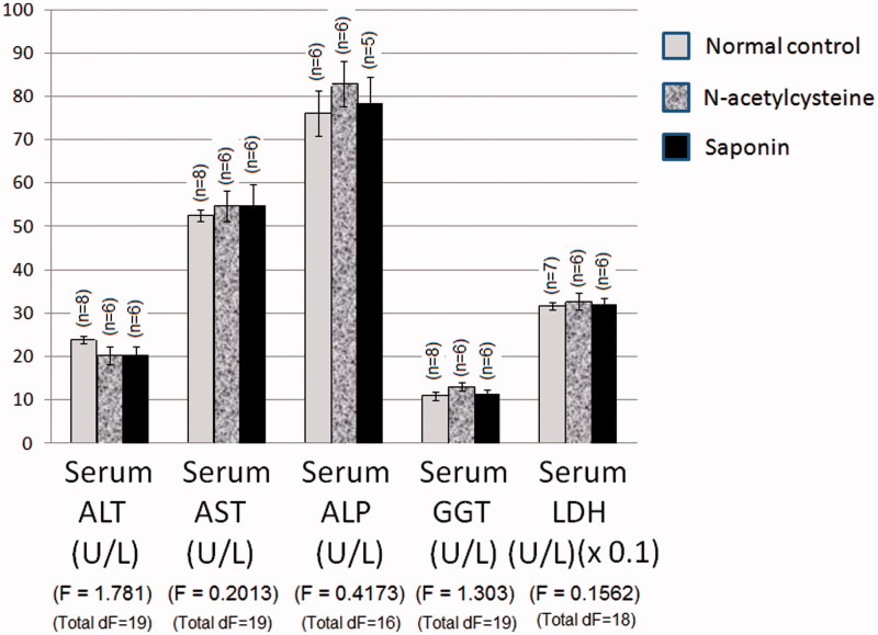
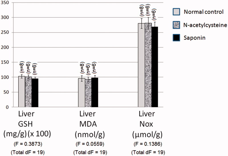
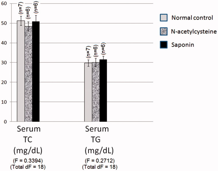
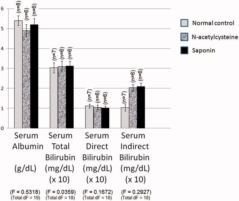
Materials and methods: Animals were divided into (six) groups, namely a normal control, an N-acetylcysteine control (300 mg/kg/day, p.o., 10 days), a saponin control (100 mg/kg/day, p.o., for 10 days), a hepatotoxicity control (two doses of ferrous sulphate, 30 mg/kg/day each, i.p., on 9th and 10th day), an N-acetylcysteine plus ferrous sulphate (standard treatment) and a saponin plus ferrous sulphate (test treatment) group. Hepatocyte integrity loss markers (serum ALT, AST, ALP, GGT and LDH), oxidative stress markers (hepatic MDA, GSH and NOx), dyslipidaemic markers (serum TC and TG) and hepatocyte functioning markers (serum bilirubin and albumin) were assessed.
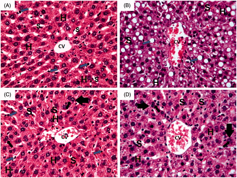
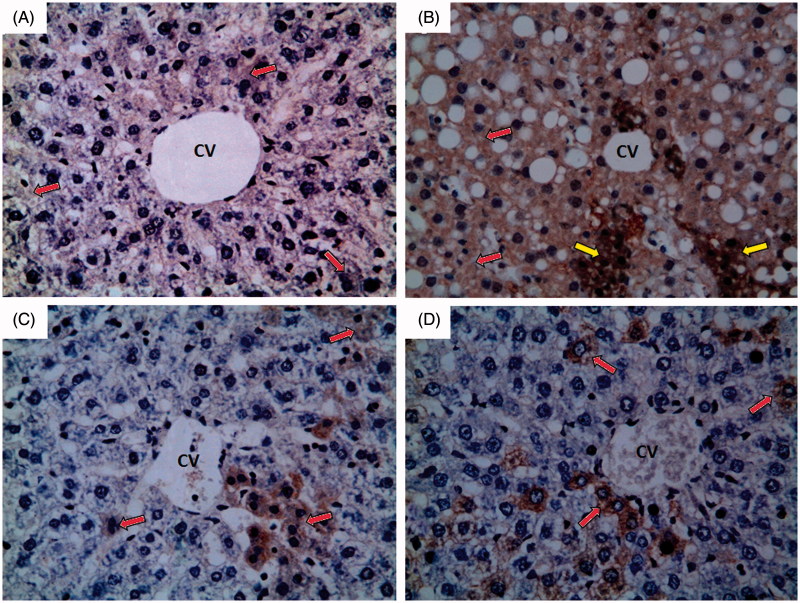
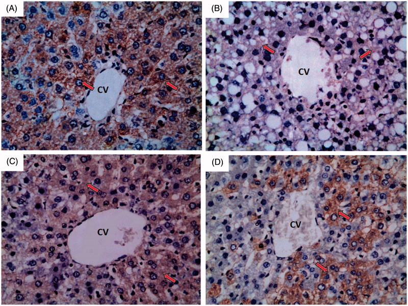
Results: Quillaja bark saponin decreased iron-induced elevation of ALT (reaching 57% of hepatotoxicity control), AST (66%), ALP (76%), GGT (60%), LDH (54%), MDA (65%), NOx (77%), TC (70%), TG (54%), and total (54%), direct (54%) and indirect (54%) bilirubin, coupled with increased GSH (219%) and albumin (159%) levels. Histopathological study strongly supported biochemical estimations, while immunohistochemical study showed marked effect on eNOS and iNOS expression.
Conclusions: Quillaja bark saponin has a good hepatoprotective effect. Amelioration of oxidative stress and suppression of NOS expression, with resultant maintenance of hepatocyte integrity and functioning, may explain this beneficial effect.
Keywords: Hepatotoxicity; N-acetylcysteine; nitric oxide synthase.
Figures
References
-
- Abdel-Fattah MM, Salama AA, Shehata BA, Ismaiel IE.. 2015a. The potential effect of the angiotensin II receptor blocker telmisartan in regulating OVA-induced airway remodeling in experimental rats. Pharmacol Rep. 67:943–951. - PubMed
-
- Abdel-Fattah MM, Messiha BA, Salama AA.. 2015b. Assessment of the mechanistic role of cinnarizine in modulating experimentally-induced bronchial asthma in rats. Pharmacology. 96:167–174. - PubMed
-
- Ahmad I, Shukla S, Kumar A, Singh BK, Kumar V, Chauhan AK, Singh D, Pandey HP, Singh C.. 2012. Biochemical and molecular mechanisms of N-acetyl cysteine and silymarin-mediated protection against maneb- and paraquat-induced hepatotoxicity in rats. Chem Biol Interact. 201:9–18. - PubMed
-
- Ali MH, Messiha BA, Abdel-Latif HA.. 2016a. Protective effect of ursodeoxycholic acid, resveratrol, and N-acetylcysteine on nonalcoholic fatty liver disease in rats. Pharm Biol. 54:1198–1208. - PubMed
-
- Ali MR, Abo-Youssef AM, Messiha BA, Khattab MM.. 2016b. Tempol and perindopril protect against lipopolysaccharide-induced cognition impairment and amyloidogenesis by modulating brain-derived neurotropic factor, neuroinflammation and oxido-nitrosative stress. Naunyn Schmiedebergs Arch Pharmacol. 389:637–356. - PubMed
MeSH terms
Substances
LinkOut - more resources
Full Text Sources
Other Literature Sources
Medical
Miscellaneous