

Occurrence of strongyloidiasis in privately owned and sheltered dogs: clinical presentation and treatment outcome
- PMID: 28728589
- PMCID: PMC5520385
- DOI: 10.1186/s13071-017-2275-5
Occurrence of strongyloidiasis in privately owned and sheltered dogs: clinical presentation and treatment outcome
Abstract
Background: The increasing number of reports of human infections by Strongyloides stercoralis from a range of European countries over the last 20 years has spurred the interest of the scientific community towards this parasite and, in particular, towards the role that infections of canine hosts may play in the epidemiology of human disease. Data on the epidemiology of canine strongyloidiasis is currently limited, most likely because of the inherent limitations of current diagnostic methods.
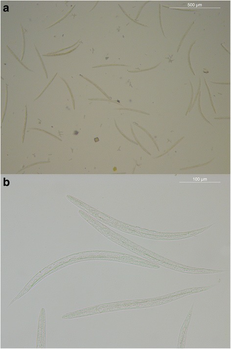
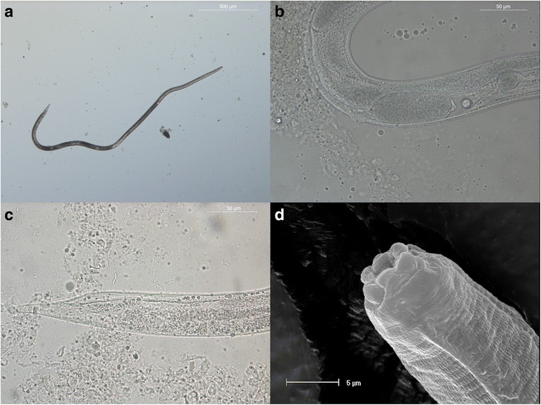
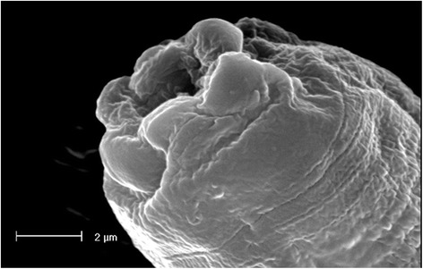
Methods: Faecal samples were collected directly from the rectal ampulla of 272 animals of varying age and both genders living in Apulia, southern Italy. Dogs included were either privately owned (n = 210), living in an urban area but with unrestricted outdoor access (Group 1), or shelter dogs (n = 62 out of ~400) hosted in a single shelter in the province of Bari in which a history of diarrhoea, weight loss, reduced appetite and respiratory symptoms had been reported (Group 2). Strongyloides stercoralis infection was diagnosed by coproscopy on direct faecal smear and via the Baermann method.
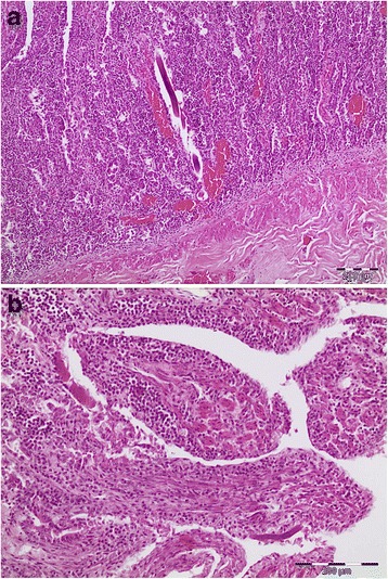
Results: Six of 272 dogs were positive for S. stercoralis at the Baermann examination; all but one were from the shelter (Group 2) and displayed gastrointestinal clinical signs. The only owned dog (Group 1) infected with S. stercoralis, but clinically healthy, had been adopted from a shelter 1 year prior to sampling. Five infected dogs were treated with fenbendazole (Panacur®, Intervet, Animal Health, 50 mg/kg, PO daily for 5 days), or with a combination of fenbendazole and moxidectin plus imidacloprid spot-on (Im/Mox; Advocate® spot-on, Bayer). Post-treatment clearance of infection was confirmed in three dogs by Baermann examination, whereas treatment failure was documented in two dogs by Baermann and/or post-mortem detection of adult parasites.
Conclusions: This study describes, for the first time, the presence of S. stercoralis infection in sheltered dogs from southern Italy. Data indicate that S. stercoralis infection may pose a concern for sheltered animals and raise questions on potential risks of infection for staff of municipal shelters in southern European countries. Given that a single course of treatment with fenbendazole, associated or not with Im/Mox spot-on, may not eliminate the infection, effective treatment protocols should be investigated and control strategies targeting the environment considered for reducing the risk of zoonotic infection.
Keywords: Clinical presentation; Dog; Faecal monitoring; Strongyloides stercoralis; Treatment; Zoonosis.
Conflict of interest statement
Ethics approval and consent to participate
Not applicable, since the procedures were part of the clinical care of symptomatic animals.
Consent for publication
Not applicable.
Competing interests
The authors declare that they have no competing interests.
Publisher’s Note
Springer Nature remains neutral with regard to jurisdictional claims in published maps and institutional affiliations.
Figures
References
-
- Viney M. Strongyloides. Parasitology. 2016;19:1–4.
-
- Nolan TJ. Canine Strongyloidiasis. In: Bowman, editor. Companion and exotic animal parasitology. Ithaca: International Veterinary Information Service (www.ivis.org); 2001.
MeSH terms
Substances
LinkOut - more resources
Full Text Sources
Other Literature Sources
