

Arousal and drug abuse
- PMID: 28729115
- PMCID: PMC5675166
- DOI: 10.1016/j.bbr.2017.07.013
Arousal and drug abuse
Abstract
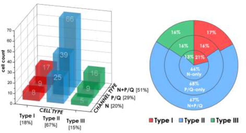
The reticular activating system (RAS) is not an amorphous region but distinct nuclei with specific membrane properties that dictate their firing during waking and sleep. The locus coeruleus and raphe nucleus fire during waking and slow wave sleep, with the pedunculopontine nucleus (PPN) firing during both waking and REM sleep, the states manifesting arousal-related EEG activity. Two important discoveries in the PPN in the last 10 years are, 1) that some PPN cells are electrically coupled, and 2) every PPN cell manifests high threshold calcium channels that allow them to oscillate at beta/gamma band frequencies. The role of arousal in drug abuse is considered here in terms of the effects of drugs of abuse on these two mechanisms. Drug abuse and the perception of withdrawal/relapse are mediated by neurobiological processes that occur only when we are awake, not when we are asleep. These relationships focus on the potential role of arousal, more specifically of RAS electrical coupling and gamma band activity, in the addictive process as well as the relapse to drug use.
Keywords: Cocaine; Connexin 36; Dopamine; Modafinil; N- and P/Q-type calcium channels; Preconscious awareness.
Copyright © 2017. Published by Elsevier B.V.
Figures
References
-
- Garcia-Rill E. Waking and the Reticular Activating System. Academic Press; New York: 2015.
-
- Takakusaki K, Shiroyama T, Yamamoto T, Kitai ST. Cholinergic and noncholinergic tegmental pedunculopontine projection neurons in rats revealed by intracellular labeling. J. Comp. Neurol. 1996;7:2353–2356. - PubMed
-
- Egan TM, North RA. Actions of acetylcholine and nicotine on rat locus coeruleus neurons in vitro. Neurosci. 1986;19:565–571. - PubMed
Publication types
MeSH terms
Grants and funding
LinkOut - more resources
Full Text Sources
Other Literature Sources
Medical
