. 2017 Jul 20;7(1):6024.
doi: 10.1038/s41598-017-06243-7.

Nucleocytoplasmic Shuttling of Histone Deacetylase 9 Controls Activity-Dependent Thalamocortical Axon Branching

Affiliations

Nucleocytoplasmic Shuttling of Histone Deacetylase 9 Controls Activity-Dependent Thalamocortical Axon Branching

Ricardo Alchini et al. Sci Rep. .

Abstract

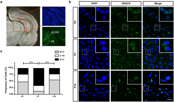
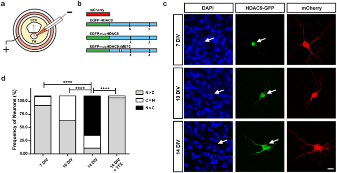
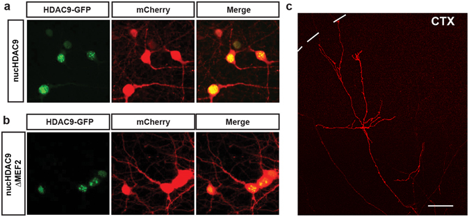
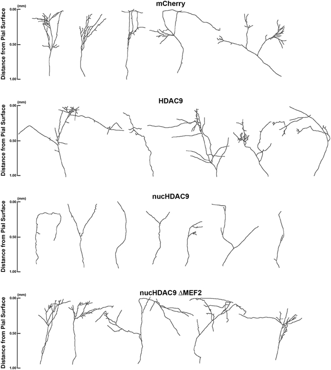
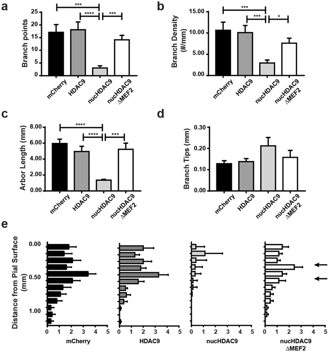
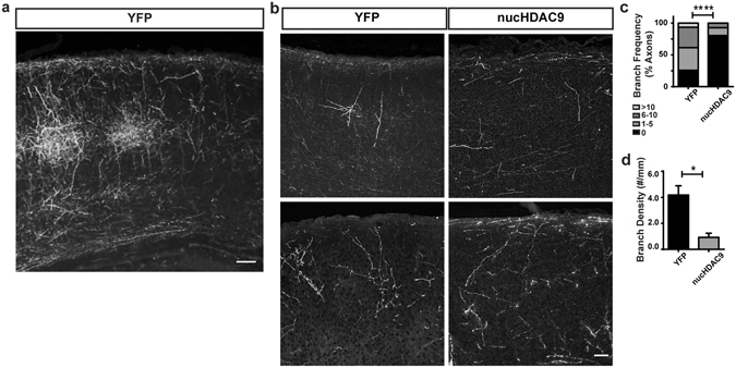
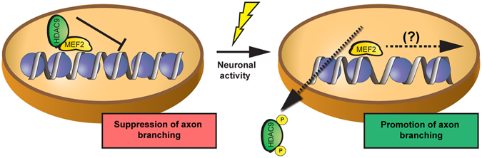
During development, thalamocortical (TC) axons form branches in an activity-dependent fashion. Here we investigated how neuronal activity is converted to molecular signals, focusing on an epigenetic mechanism involving histone deacetylases (HDACs). Immunohistochemistry demonstrated that HDAC9 was translocated from the nucleus to the cytoplasm of thalamic cells during the first postnatal week in rats. In organotypic co-cultures of the thalamus and cortex, fluorescent protein-tagged HDAC9 also exhibited nuclueocytoplasmic translocation in thalamic cells during culturing, which was reversed by tetrodotoxin treatment. Transfection with a mutant HDAC9 that interferes with the translocation markedly decreased TC axon branching in the culture. Similarly, TC axon branching was significantly decreased by the mutant HDAC9 gene transfer in vivo. However, axonal branching was restored by disrupting the interaction between HDAC9 and myocyte-specific enhancer factor 2 (MEF2). Taken together, the present results demonstrate that the nucleocytoplasmic translocation of HDAC9 plays a critical role in activity-dependent TC axon branching by affecting transcriptional regulation and downstream signaling pathways.


Conflict of interest statement

The authors declare that they have no competing interests.

Figures

Figure 1
Figure 1
HDAC9 translocation in the developing rat thalamus. (a) Locations of dLGN and VB are shown in a coronal section. (b) P0, P7 and P14 rat thalami were analyzed after immunohistochemistry against HDAC9. HDAC9 export from the nucleus can be observed by the end of the first postnatal week. Scale bar: 35 μm Green: HDAC9. Blue: DAPI. (c) Distribution of the cells according to the ratio of HDAC9 signal intensity in the cytoplasm normalized to its nuclear intensity. Cells in which the cytoplasm/nuclear signal ratio was below 0.9 were considered as having a nuclear signal (N > C). Cells that the average signal in the nucleus and the cytoplasm did not differ in more than 10% (i.e. ratios between 0.9 and 1.1) were categorize as N = C. Cells in which the signals in the cytoplasm were considerably higher than in the nucleus (ratios above 1.1) were categorized as N < C. N = 958, 2029 and 808 cells from 3 different animals for P0, P7 and P14, respectively. Statistical analysis by overall Chi-square (Chi = 898.7, with 4 degrees of freedom, p < 0.0001) followed by pairwise Chi-square with Bonferroni correction. ***p < 0.001.
Figure 2
Figure 2
Subcellular localization of HDAC9-EGFP fusion protein in TC organotypic co-culture preparations. (a) Experimental set-up of the co-culture preparation and electroporation into thalamic cells. (b) Schematic representation of the plasmids used in the present study (see the methods). (c) Representative thalamic neurons at 7, 10 and 14 DIV. HDAC9-EGFP distribution in the cells can be compared to the cytoplasmic protein mCherry and the nuclear staining of DAPI. Arrows refer to the position of the same cell at a given time point. Scale bar 10 μm. (d) Quantification of subcellular localization of HDAC9 was expressed as the ratio of transfected cells to illustrate HDAC9-EGFP signal concentrated predominantly in the nucleus (N > C), equally in the nucleus and the cytoplasm (N = C) or predominantly in the cytoplasm (N < C). Statistical analysis by overall Chi-square (Chi = 158, with 6 degrees of freedom, p < 0.000001) followed by pairwise Chi-square with Bonferroni correction. ****p < 0.0001.
Figure 3
Figure 3
Subcellular localization of HDAC9 mutants in thalamic cells and TC axon branching in vitro. Both of nucHDAC9-EGFP (a) and nucHDAC9ΔMEF2-EGFP (b) were distributed in the nuclei of thalamic cells at 14 DIV, while co-expressed mCherry was distributed in cell bodies and dendrites. Green: EGFP signal from the fusion protein. Red: mCherry. Scale bar, 5 μm. (c) Representative mCherry-labeled axon in the cortical explant, which elongated from the transfected thalamic cell. Interrupted line indicates the pial surface of the cortical explant. Scale bar 100 μm.
Figure 4
Figure 4
Disruption of HDAC9 translocation and MEF2 interaction affect TC axon branching. Representative tracings of control and mutant HDAC9 transfected axons. Thalamic cells were electroporated with only mCherry to observe control TC axon branching. To examine the effect of the HDAC9 mutants, mCherry was co-transfected with either of HDAC9, nucHDAC9, or nucHDAC9ΔMEF2.
Figure 5
Figure 5
Nucleocytoplasmic translocation of HDAC9 is required for TC axon branching via MEF2 regulation. (a) Average number of branch points per axon. Thalamic cells were electroporated with only mCherry, HDAC9 co-electroporated with mCherry, nucHDAC9 co-electroporated with mCherry, and nucHDAC9ΔMEF2 co-electroporated with mCherry. N = 12, 17, 18 and 14 axons from 4–6 co-cultures for each condition, respectively. (b) Branch density per axon. (c) Total branch length per axon. (d) Branch tip length. Statistical analysis by Kruskal-Wallis test with Dunn correction comparing the 4 columns. *p < 0.05. ***p < 0.001. ****p < 0.0001. e. Laminar distribution of branching points according to their distance from the pial surface. Arrows indicate presumed layer 4. Statistical analysis by two-way ANOVA for whole column effects with Tukey’s multiple comparisons test confirmed a significant difference in laminar specificity between nucHDAC9 and control (p < 0.0001), while no significant difference was found between nucHDAC9ΔMEF2 and control or wild-type HDAC9.
Figure 6
Figure 6
HDAC9 nuclear export enhances axon branching in vivo. (a) EYFP-labeled TC axonal projection in the P7 SSp cortex of an animal whose thalamus was electroporated with EYFP plasmid. Coronal section of approximately 60 μm thickness. Scale bar 100 μm. (b) Representative TC axon fragments from control (EYFP-electroporated) and nucHDAC9/mCherry co-electroporated neurons. Coronal sections of 20 μm thickness. Scale bar 50 μm. Statistical analysis by Chi-square (Chi = 22.3, with 3 degrees of freedom), p < 0.0001. (c) Distribution of axon segments according to their branch number. (d) Average branch density. Statistical analysis by Student t-test. *p < 0.05.
Figure 7
Figure 7
Working model of HDAC9 function in axon branch formation. HDAC9 is confined to the nucleus, where it can bind to transcription factors such as MEF2 and represses axon branching. Neuronal activity induces HDAC9 export to the cytoplasm, which promotes axon branching.

References

    1. Shatz CJ, Stryker MP. Prenatal tetrodotoxin infusion blocks segregation of retinogeniculate afferents. Science. 1988;242:87–89. doi: 10.1126/science.3175636. - DOI - PubMed
    1. Hubel DH, Wiesel TN, LeVay S. Plasticity of ocular dominance columns in monkey striate cortex. Philos Trans R Soc Lond B Biol Sci. 1977;278:377–409. doi: 10.1098/rstb.1977.0050. - DOI - PubMed
    1. Antonini A, Stryker MP. Rapid remodeling of axonal arbors in the visual cortex. Science. 1993;260:1819–1821. doi: 10.1126/science.8511592. - DOI - PubMed
    1. Agmon A, Yang LT, O’Dowd DK, Jones EG. Organized growth of thalamocortical axons from the deep tier of terminations into layer IV of developing mouse barrel cortex. J Neurosci. 1993;13:5365–5382. - PMC - PubMed
    1. Rebsam A, Seif I, Gaspar P. Refinement of thalamocortical arbors and emergence of barrel domains in the primary somatosensory cortex: a study of normal and monoamine oxidase a knock-out mice. J Neurosci. 2002;22:8541–8552. - PMC - PubMed

Publication types

Substances

LinkOut - more resources