Gastric Hemangioma Treated with Argon Plasma Coagulation in a Newborn Infant
- PMID: 28730139
- PMCID: PMC5517381
- DOI: 10.5223/pghn.2017.20.2.134
Gastric Hemangioma Treated with Argon Plasma Coagulation in a Newborn Infant
Abstract
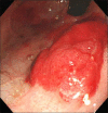
Gastric hemangioma in the neonatal period is a very rare cause of upper gastrointestinal bleeding. We present a case of hemangioma limited to the gastric cavity in a 10-day-old infant. A huge, erythematous mass with bleeding was observed on the lesser curvature side of the upper part of the stomach. Surgical resection was ruled out because the location of the lesion was too close to the gastroesophageal junction. Medical treatment with intravenous H2 blockers, octreotide, packed red blood cell infusions, local epinephrine injection at the lesion site, application of hemoclip, and gel-form embolization of the left gastric artery did not significantly alter the transfusion requirement. Hemostasis was achieved with endoscopic argon plasma coagulation (APC). After two sessions of APC, complete removal of the lesion was achieved. APC was a simple, safe and effective tool for hemostasis and the ablation of gastric hemangioma without significant complications.
Keywords: Argon plasma coagulation; Hemangioma; Neonate; Stomach.
Figures


References
-
- Menon P, Rao KL, Bhasin S, Vanitha V, Thapa BR, Lal A, et al. Giant isolated cavernous hemangioma of the stomach. J Pediatr Surg. 2007;42:747–749. - PubMed
-
- Bamanikar AA, Diwan AG, Benoj D. Gastric hemangioma: an unusual cause of upper gastrointestinal bleed. Indian J Gastroenterol. 2004;23:113–114. - PubMed
-
- Fishman SJ, Burrows PE, Leichtner AM, Mulliken JB. Gastrointestinal manifestations of vascular anomalies in childhood: varied etiologies require multiple therapeutic modalities. J Pediatr Surg. 1998;33:1163–1167. - PubMed
-
- López-Gutiérrez JC. Hemangiomas and vascular malformations of the stomach. J Pediatr Surg. 2007;42:1634–1635. - PubMed
-
- Nagaya M, Kato J, Niimi N, Tanaka S, Akiyoshi K, Tanaka T. Isolated cavernous hemangioma of the stomach in a neonate. J Pediatr Surg. 1998;33:653–654. - PubMed
Publication types
LinkOut - more resources
Full Text Sources
Other Literature Sources

