Review
. 2018 Jan;75(2):261-273.
doi: 10.1007/s00018-017-2599-5. Epub 2017 Jul 20.

Regulation of class V myosin

Affiliations
Review

Regulation of class V myosin

Ning Zhang et al. Cell Mol Life Sci. 2018 Jan.

Abstract

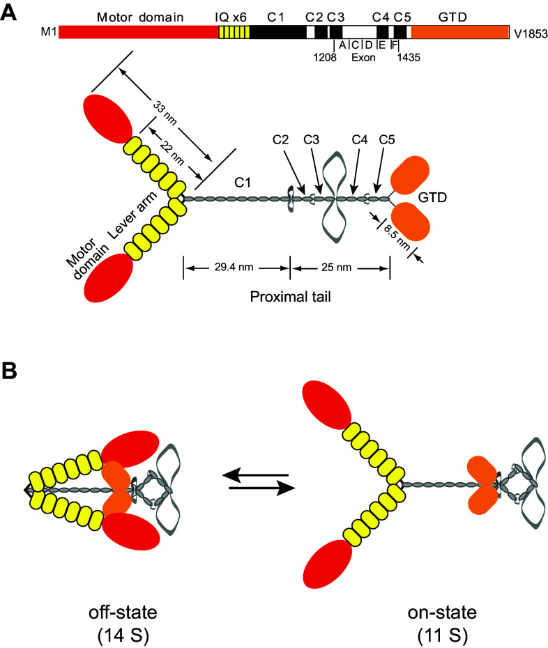
Class V myosin (myosin-5) is a molecular motor that functions as an organelle transporter. The activation of myosin-5's motor function has long been known to be associated with a transition from the folded conformation in the off-state to the extended conformation in the on-state, but only recently have we begun to understand the underlying mechanism. The globular tail domain (GTD) of myosin-5 has been identified as the inhibitory domain and has recently been shown to function as a dimer in regulating the motor function. The folded off-state of myosin-5 is stabilized by multiple intramolecular interactions, including head-GTD interactions, GTD-GTD interactions, and interactions between the GTD and the C-terminus of the first coiled-coil segment. Any cellular factor that affects these intramolecular interactions and thus the stability of the folded conformation of myosin-5 would be expected to regulate myosin-5 motor function. Both the adaptor proteins of myosin-5 and Ca2+ are potential regulators of myosin-5 motor function, because they can destabilize its folded conformation. A combination of these regulators provides a versatile scheme in regulating myosin-5 motor function in the cell.

Keywords: Actin; Allosteric regulation; Calmodulin; Molecular motor; Myosin-5.


Conflict of interest statement

The authors declare that they have no conflicts of interest with the contents of this article.

Figures

Fig. 1
Fig. 1
Structural arrangement and regulation of Myo5a. a Domain arrangement (top) and the predicted structure of Myo5a (bottom). At the amino terminus is the motor domain containing the ATP- and actin-binding sites. The motor domain is followed by the lever arm that consists of six IQ motifs (IQ × 6). The consensus sequence is IQXXXRGXXXR, which act as the binding sites for CaM or CaM-like light chains. The next ~500 amino acids are predicted to form five coiled-coils (C1C5) separated by four flexible regions. The last ~400 amino acids form a globular tail domain (GTD). The regions encoded by the exons (A–F) discussed in the review are indicated. The lengths of the proximal tail are calculated based on the length of the predicted coiled-coils, and those of the motor domain and the GTD are estimated from the crystal structures. b Tail-inhibition model for the regulation of Myo5a motor function. The off-state of Myo5a is in the 14S folded conformation in which the two GTDs associate with the C-terminus of the coiled-coil-1 and bind to the motor domains, thus forming an isosceles triangle conformation. The interruption of the head–GTD interaction (either by adaptor protein binding or elevation of Ca2+) activates Myo5a to form the 11S extended conformation. This figure was modified from [21]. Note: the extended conformation of Myo5a in (a) is different from that in (b), in which the GTD binds to the C-terminus of coiled-coil-1
Fig. 2
Fig. 2
Structures of the head and the GTD of myosin-5. a Structure of Myo5a containing the motor domain and IQ1 in complex with ELC (an equivalent of apo-CaM) (PDB code 1W7J). The motor domain contains the nucleotide-binding site and the actin-binding region. The putative GTD-binding site is composed of the SH3-linker (P65–L77), loop-136 (G129–D136), the converter, the proximal portion of lever arm (G754–D765), and the C-lobe of CaM. This figure was modified from [93]. b GTD structure. Top the structure of Myo5a–GTD in complex of Mlph–GTBM (PDB code 4LX1). Bottom the structure of Myo5b–GTD (PDB code 4J5M) dimer. The binding sites for the motor domain and for two other adaptor proteins, Rab11 and RILPL2–RH1, are shown. Both the conserved basic residues K1706/1779 (shown in spheres) and H11–H12 loop (shown in red) are essential for the head interaction. Sub-1 and -2, subdomain-1 and -2 of the GTD; Mlph–GTBM, the GTD-binding motif of Mlph. This figure was modified from [94]
Fig. 3
Fig. 3
Structural model of the off-state of Myo5a. A diagram of the triangular structure of the off-state of Myo5a is shown in the upper left panel. The model of the motor domain and the first two IQ motifs domain of Myo5a was created from the motor domain of chicken Myo5a in a post-rigor state (PDB code 1W7J) and the first two IQ motifs domain of mouse Myo5a (PDB code 2IX7) by superposing the backbone of R767–I773. The model of the interaction between the head and the GTD was created from the Myo5a head in a post-rigor state (above) and the Myo5b–GTD dimer (PDB code 4J5M) by positioning D136 in the motor domain and K1706/K1779 in Myo5b–GTD within a distance of ~4 Å and ensuring to avoid the collision of the structures. Sub-1 and -2, subdomain-1 and -2 of the GTD; CaM-1 and -2, the CaM bound to IQ1 and IQ2 of Myo5a
Fig. 4
Fig. 4
Strategies for activating myosin-5 motor function. (a) Removal of GTD inhibition, and thus activation of motor activity, could occur upon binding of cargo to the GTD. (b) Binding of cargo to the tail (including the GTD and/or the proximal tail portion) could be insufficient to remove the GTD inhibition and further events such as (c) other adaptor proteins or (d) Ca2+, might be involved. (e) Ca2+ might induce the free myosin-5 to transform from the off-state to the on-state and then bind to the cargo (f). It should be noted that the role of Ca2+ as a major physiological regulatory mechanism for myosin-5 remains controversial

References

    1. Odronitz F, Kollmar M. Drawing the tree of eukaryotic life based on the analysis of 2269 manually annotated myosins from 328 species. Genome Biol. 2007;8:R196. doi: 10.1186/gb-2007-8-9-r196. - DOI - PMC - PubMed
    1. Foth BJ, Goedecke MC, Soldati D. From the cover: new insights into myosin evolution and classification. Proc Natl Acad Sci USA. 2006;103:3681–3686. doi: 10.1073/pnas.0506307103. - DOI - PMC - PubMed
    1. Heissler SM, Sellers JR. Kinetic adaptations of myosins for their diverse cellular functions. Traffic. 2016;17:839–859. doi: 10.1111/tra.12388. - DOI - PMC - PubMed
    1. Woolner S, Bement WM. Unconventional myosins acting unconventionally. Trends Cell Biol. 2009;19:245–252. doi: 10.1016/j.tcb.2009.03.003. - DOI - PMC - PubMed
    1. Heissler SM, Sellers JR. Various themes of myosin regulation. J Mol Biol. 2016;428:1927–1946. doi: 10.1016/j.jmb.2016.01.022. - DOI - PMC - PubMed

LinkOut - more resources