Review
. 2017 Aug 9;117(15):10474-10501.
doi: 10.1021/acs.chemrev.7b00117. Epub 2017 Jul 21.

The Metal Drives the Chemistry: Dual Functions of Acireductone Dioxygenase

Affiliations
Review

The Metal Drives the Chemistry: Dual Functions of Acireductone Dioxygenase

Aditi R Deshpande et al. Chem Rev. .

Abstract

Acireductone dioxygenase (ARD) from the methionine salvage pathway (MSP) is a unique enzyme that exhibits dual chemistry determined solely by the identity of the divalent transition-metal ion (Fe2+ or Ni2+) in the active site. The Fe2+-containing isozyme catalyzes the on-pathway reaction using substrates 1,2-dihydroxy-3-keto-5-methylthiopent-1-ene (acireductone) and dioxygen to generate formate and the ketoacid precursor of methionine, 2-keto-4-methylthiobutyrate, whereas the Ni2+-containing isozyme catalyzes an off-pathway shunt with the same substrates, generating methylthiopropionate, carbon monoxide, and formate. The dual chemistry of ARD was originally discovered in the bacterium Klebsiella oxytoca, but it has recently been shown that mammalian ARD enzymes (mouse and human) are also capable of catalyzing metal-dependent dual chemistry in vitro. This is particularly interesting, since carbon monoxide, one of the products of off-pathway reaction, has been identified as an antiapoptotic molecule in mammals. In addition, several biochemical and genetic studies have indicated an inhibitory role of human ARD in cancer. This comprehensive review describes the biochemical and structural characterization of the ARD family, the proposed experimental and theoretical approaches to establishing mechanisms for the dual chemistry, insights into the mechanism based on comparison with structurally and functionally similar enzymes, and the applications of this research to the field of artificial metalloenzymes and synthetic biology.


Conflict of interest statement

Notes

The authors declare no competing financial interest.

Figures

Figure 1
Figure 1. β-barrel structures in cupins
a) Oxalate oxidase (PDB ID: 2ET1), b) Clavaminic acid synthase (PDB ID: 1GVG) is an example of a ketoglutarate-dependent oxidase, c) homoprotocatechuate 2,3-dioxygenase (PDB ID: 3OJT) a non-heme iron-dependent dioxygenase, d) Oxalate decarboxylase (PDB ID: 1L3J). The beta sheets are colored red and the helixes are blue.
Figure 2
Figure 2. The Methionine Salvage Pathway in Klebsiella oxytoca
Reproduced with permission from Ref. . Copyright 2016 American Chemical Society.
Figure 3
Figure 3. Structures of Fe- and Ni-bound KoARD
Secondary structural features are labeled corresponding to primary sequence: A: Ala2-Phe6, B: Ser14-Ser18, C: Ala21-Lys31, D: Val33-Thr49, E: Ala50-Ala60, F: Ile61-Gly69, G: Ser72-Ile76, H: Pro83-Asn94, I: Glu102-Glu108, J: Leu112-His116, K: Val121-Leu125, L: Leu131-Val134, M: His140-Met144, N: Thr151-Phe156, O: Asn158-Gly161, P: Thr162-Ile163, Q: Ile171-Tyr175.
Figure 4
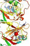
Figure 4. Active sites of Ni- and Fe-bound KoARD
Close-up views of the actives sites of Ni-KoARD (1ZRR, top) and Fe-KoARD (2HJI, bottom). Secondary structures O and P in the Ni enzyme are disordered in the Fe enzyme and are not shown in the bottom panel. The Ni view is scaled slightly smaller than the Fe view so that relevant features can be displayed.
Figure 5
Figure 5. Crystal structures of ARD enzymes from B. anthracis, H. sapiens and M. musculus
Coordinated metal ions are shown as red spheres. a) BaARD (PDB ID: 4QGM), b) HsARD (PDB ID: 4QGN), c) MmARD (PDB ID: 1VR3)
Figure 6
Figure 6. X-ray crystal structure of Ni-MmARD (PDB ID: 5I91)
a) The crystallographic structure of Ni-MmARD. The Nickel atom is shown as a green sphere and its protein ligands His88, His90, His133 and Glu94 represented as sticks and two water molecules are shown as red dots. b) Stereo view of nickel binding in the MmARD active site. Glu94 and His88 provide the axial ligands, with His90, His133 and two water/hydroxide ligands occupying equatorial positions, yielding an octahedral coordination geometry. The metal-ligand distances (in Å) are show as red dotted lines. (Reproduced with permission from Ref . Copyright 2016 American Chemical Society).
Figure 7
Figure 7. The active site of Ni-MmARD showing substrate-analog and product binding
The Ni atom is shown as a green sphere, the ligands and active site residues are shown as sticks and waters/hydroxides are shown as small red spheres. Hydrogen bonding distances are shown (in Å) as red dotted lines. Hydrophobic residues in the active site are shown in yellow. a) Stereo view of KMTB bound to Ni-MmARD. KMTB is within hydrogen bonding distance of Arg96 and the two water molecules ligated to Ni. The residues Phe84, Phe105, Phe135, Ala145, Val143 and Ile98 interact with the alkyl group of KMTB. b) Stereo view of D-Lactic acid (D-LA) bound to Ni-MmARD. D-LA replaces both equatorial water ligands, coordinating in a bidentate manner with Ni2+ via the carboxylate and hydroxyl oxygens. D-LA is within hydrogen bonding distance of Arg96. Residues Phe84, Phe105 and Phe135 interact with the alkyl group of D-LA. (Adapted from Ref . Copyright 2016 American Chemical Society).
Figure 8
Figure 8. Sequence alignment of ARD homologs using ClustalOmega
The metal binding residues His88, His90, Glu94 and His133 are colored blue, the hydrophobic residues Phe84, Phe105, Phe135 and Ala145 found to be interacting with KMTB are colored red. Arg96 and Arg147 are colored green. Phe149 and Trp155 may be involved in conformational gating are shown in magenta. (All residue numbers refer to the MmARD sequence). The gap in the eukaryotic sequences (corresponding to the sequence APTAETVIAA in Klebsiella is due to a shortened flexible loop D between strand C and helix E (Fig. 3).
Figure 9
Figure 9. Surface representation of MmARD showing residues likely involved in conformational gating
KMTB, Phe149 and Trp155 are shown in stick representation, Ni is shown as a green sphere and the water molecules shown as red spheres. Trp155 resides in the loop region preceding the long C-terminal helix, and may be involved in conformational changes upon substrate binding. Phe149 may be involved in the assisting Trp155 in conformational gating. a) Overall structure, b) Overall structure showing the C-terminal helix and the loop preceding it in a cartoon representation, c) Active site structure showing Trp155, d) Active site structure showing Trp155 & Phe149.
Figure 10
Figure 10. Solution NMR data of Mn2+, Fe2+, Co2+ and Ni2+-bound HsARD isozymes
Left: Upfield region of 800 MHz 1H NMR spectra of Mn-HsARD (pink), Fe-HsARD (red), Co-HsARD (blue), and Ni-HsARD (green) showing methyl resonances of amino acid side chains ring current-shifted by nearby aromatic residues. The different spectral patterns indicate differences in side chain packing in hydrophobic cores. Right: Overlay of the 2D 1H, 15N HSQC spectra of Mn-HsARD (pink), Co-HsARD (blue), Fe-HsARD (red) and Ni-HsARD (green). Different shift patterns indicate differences in local hydrogen bonding and structure. All spectra were obtained at 25 °C, pH 7.0 at 800 MHz 1H observe frequency. Reproduced with permission from Ref. . Copyright 2017 Oxford University Press.
Scheme 1
Scheme 1
Synthetic route to ARD substrates (Adapted from Ref. 46)
Scheme 2
Scheme 2
Model reactions showing feasibility of peroxy intermediates in Ni KoARD activity (Adapted from Ref. 46).
Scheme 3
Scheme 3
Proposed mechanism for inactivation of ARD via a radical intermediate generated by 1-electron transfer from substrate analog 5b (Adapted from Ref. 46).
Scheme 4
Scheme 4. Proposed mechanisms of Ni-ARD and Fe-ARD using the chelate hypothesis
The results of incorporation of 18O and 14C labeling-studies are indicated by the red O atoms and the asterisk. Reproduced with permission from Ref . Copyright 2016 American Chemical Society.
Scheme 5
Scheme 5. Proposed mechanism of Ni-ARD chemistry using small molecule model of Ni-KoARD active site
(Figure adapted from Ref. 94)
Scheme 6
Scheme 6. Proposed mechanism of Fe-ARD chemistry using small molecule model of Fe-KoARD active site
(Figure adapted from Ref. 94)
Scheme 7
Scheme 7. Proposed mechanism of Ni-KoARD and Fe-KoARD using computational studies
(Adapted from Ref. 19)
Scheme 8
Scheme 8. Reactions catalyzed by oxalate decarboxylase (OxDC), oxalate oxidase (OxOx) and quercetinase (QueD)
Scheme 9
Scheme 9. Proposed catalytic mechanism of oxalate oxidase
The protein ligands, His88, His90, His135, and Glu95 for Mn2+ are not shown. (Figure adapted from Ref. 67)
Scheme 10
Scheme 10. Proposed catalytic mechanism of Streptomyces sp. FLA QueD
The protein ligands, His69, His71 and His115 for Ni2+ are not shown. (Figure adapted from Ref. 65)
Scheme 11
Scheme 11. Proposed catalytic mechanism of homoprotocatechuate 2,3-dioxygenase which is an extradiol dioxygenase
(R = -CH2COOH) (Figure adapted from Ref. 9)
Scheme 12
Scheme 12. Ternary enzyme-substrate-dioxygen complexes for HPCD, HAD and HGDO observed by X-ray crystallography

Similar articles

Cited by

References

    1. Dai Y, Wensink PC, Abeles RH. One protein, two enzymes. J Biol Chem. 1999;274(3):1193–1195. - PubMed
    1. Wray JW, Abeles RH. The methionine salvage pathway in Klebsiella pneumoniae and rat liver. Identification and characterization of two novel dioxygenases. J Biol Chem. 1995;270(7):3147–3153. - PubMed
    1. Deshpande AR, Wagenpfeil K, Pochapsky TC, Petsko GA, Ringe D. Metal-Dependent Function of a Mammalian Acireductone Dioxygenase. Biochemistry. 2016;55(9):1398–1407. - PMC - PubMed
    1. Deshpande AR, Pochapsky TC, Petsko GA, Ringe D. Dual chemistry catalyzed by human acireductone dioxygenase. Protein Eng Des Sel. 2017;30(3):197–204. - PMC - PubMed
    1. Yeh CT, Lai HY, Chen TC, Chu CM, Liaw YF. Identification of a hepatic factor capable of supporting hepatitis C virus replication in a nonpermissive cell line. J Virol. 2001;75(22):11017–11024. - PMC - PubMed