. 2017 Jul 21;12(7):e0181656.
doi: 10.1371/journal.pone.0181656. eCollection 2017.

The exported chaperone Hsp70-x supports virulence functions for Plasmodium falciparum blood stage parasites

Affiliations

The exported chaperone Hsp70-x supports virulence functions for Plasmodium falciparum blood stage parasites

Sarah C Charnaud et al. PLoS One. .

Abstract

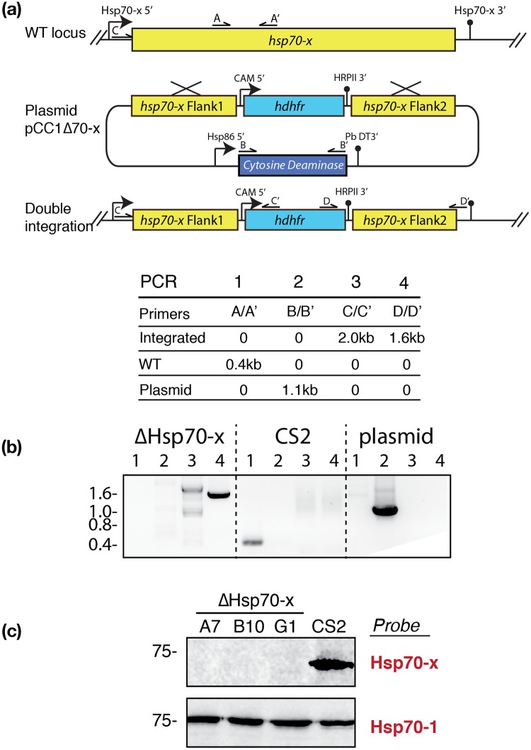
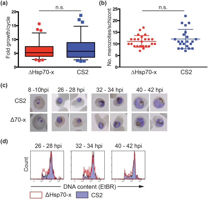
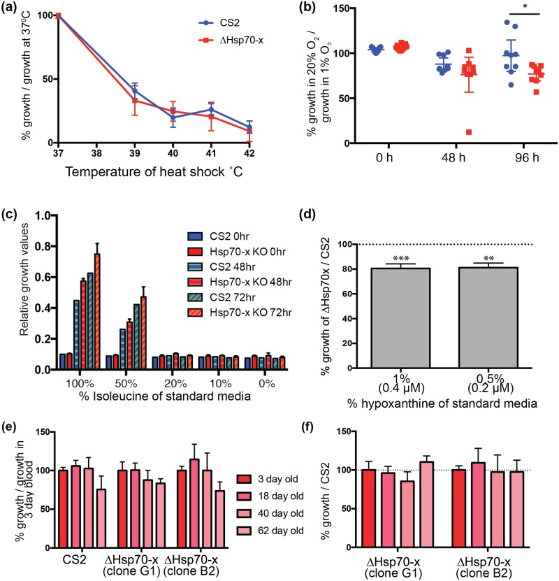
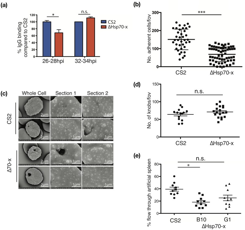
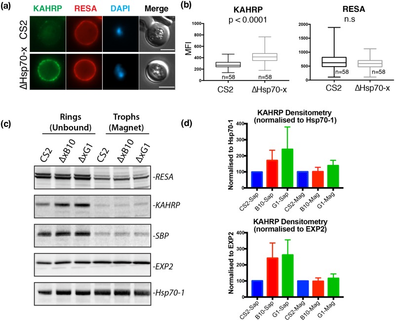
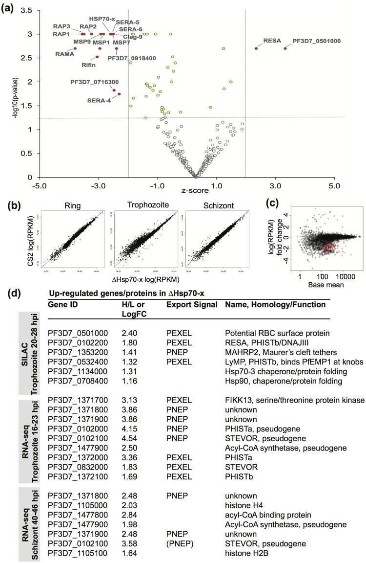
Malaria is caused by five different Plasmodium spp. in humans each of which modifies the host erythrocyte to survive and replicate. The two main causes of malaria, P. falciparum and P. vivax, differ in their ability to cause severe disease, mainly due to differences in the cytoadhesion of infected erythrocytes (IE) in the microvasculature. Cytoadhesion of P. falciparum in the brain leads to a large number of deaths each year and is a consequence of exported parasite proteins, some of which modify the erythrocyte cytoskeleton while others such as PfEMP1 project onto the erythrocyte surface where they bind to endothelial cells. Here we investigate the effects of knocking out an exported Hsp70-type chaperone termed Hsp70-x that is present in P. falciparum but not P. vivax. Although the growth of Δhsp70-x parasites was unaffected, the export of PfEMP1 cytoadherence proteins was delayed and Δhsp70-x IE had reduced adhesion. The Δhsp70-x IE were also more rigid than wild-type controls indicating changes in the way the parasites modified their host erythrocyte. To investigate the cause of this, transcriptional and translational changes in exported and chaperone proteins were monitored and some changes were observed. We propose that PfHsp70-x is not essential for survival in vitro, but may be required for the efficient export and functioning of some P. falciparum exported proteins.


Conflict of interest statement

Competing Interests: The authors have declared that no competing interests exist.

Figures

Fig 1
Fig 1. hsp70-x can be knocked out and is not essential for parasite survival.
(a) Diagram of plasmid construct indicating double homologous recombination event to replace hsp70-x with a hdhfr drug resistance cassette. Negative selection pressure to remove parasites carrying non-integrated plasmid is provided by the cytosine deaminase. The binding sites of PCR primers used to confirm gene replacement are indicated. (b) (Top) Table of PCR primer combinations and expected DNA product sizes resulting from amplification the native hsp70-x locus, the hdhfr replacement locus and plasmid construct are shown. (Bottom) Agarose gel of PCR products from one Δhsp70-x clone, the parental (CS2) parasites and plasmid only controls indicate the hsp70-x gene has been deleted by double cross over integration. (c) Western blot probed with rabbit anti-Hsp70-x confirms three separate ΔHsp70-x clones no longer express Hsp70-x, but retain the closely related cytoplasmic Hsp70-1.
Fig 2
Fig 2. Deletion of hsp70-x does not affect parasite growth in the blood stage cell cycle.
(a) Measurement of the fold increase cell proliferation per blood stage cycle of ΔHsp70-x and CS2 parasites as assessed by parasite lactate dehydrogenase (LDH) activity indicates the growth of ΔHsp70-x parasites was not significantly (n.s.) slower than CS2. Mann-Whitney p = 0.7, n = 36. (b) Light microscopy counts of the mean number of merozoites per schizont in ΔHsp70-x and CS2 parasite lines indicated there was no significant difference. (c) Light microscopy images of four hour synchronized Giemsa stained parasites indicating both lines were morphologically similar. (d) Measurement of the DNA content of ethidium bromide (EtBR) stained ΔHsp70-x and CS2 IE by flow cytometry indicates similar DNA content in the majority of cells at each time point. Parasites were synchronized to a four-hour window and the red trace indicates ΔHsp70-x DNA content compared to CS2 in purple.
Fig 3
Fig 3. ΔHsp70-x parasites generally resist stress as well as CS2.
(a) Growth of ΔHsp70-x and CS2 parasites for 1.5 cell cycles after heat shock treatment of ring stage parasites indicated that growth of the mutant ΔHsp70-x and CS2 were similarly reduced. The ring-stage parasites were heat shocked at indicated temperatures for six hours and the percentage growth relative to parasite growth at 37°C is shown. The parasite lactate dehydrogenase (LDH) assay was used to measured growth and was performed twice in triplicate. (b) Percentage of blood stage parasite growth in 5% CO2 in normal air (20% O2) as a proxy for oxidative stress, relative to non-stressed cultures in 5% CO2, 1% O2. Both CS2 and ΔHsp70-x have reduced growth in oxidative stress conditions at 48 h with ΔHsp70-x not recovering as well as some of the CS2 replicates at 96 h. Growth was measured by LDH assay and performed in triplicate, showing the mean with 95% CI * p = 0.02. (c) Growth in media with reduced levels of isoleucine as measured by LDH assay at 0, 48 and 72 h after start of growth period, n = 2. For most isoleucine concentrations there was no difference between the ΔHsp70-x and CS2 parasites. (d) With hypoxanthine reduced to 1% (0.4 μM) and 0.5% (0.2 μM) of standard RPMI culture conditions, the growth of ΔHsp70-x was slightly reduced compared to CS2 as indicated by the dashed line at 100% (n = 3). Growth was measured by LDH activity after two cycles and was performed with two clones of ΔHsp70-x whose data were combined because they were not significantly different to each other. T-test comparing ΔHsp70-x growth to CS2 p = *** > 0.0001, ** 0.0008. (e) Growth of parasite lines in different aged erythrocytes as measured by LDH activity after 2.5 cell cycles indicates both parasite lines grew less well in older erythrocytes. The erythrocytes were stored at 4°C for the indicated number of days prior to start of the assay. Parasite LDH was measured five days after the start of the assay and is reported as % growth compared to parasites grown in 3 day old blood, n = 6. (f) Data from (e) showing the growth of ΔHsp70-x clones compared to control CS2 parasites normalised to 100% for each erythrocyte age group indicates no difference.
Fig 4
Fig 4. ΔHsp70-x parasites export PfEMP1 less efficiently, bind less well under flow and have altered cell rigidity.
(a) Recognition of surface exposed PfEMP1 by human immune sera indicates there is a delay in the deployment of PfEMP1 on the surface of young (26–28 hpi) but not older (32–34 hpi) ΔHsp70-x infected erythrocytes compared to CS2. Flow cytometry was used to measure the mean fluorescence intensity of PfEMP1 on synchronized parasites and was performed in triplicate. T-test *p = 0.04, n = 2. (b) Cytoadhesion of infected cells at 32–34 hpi under a 0.05 Pa flow rate to simulate microvasculature conditions indicates that ΔHsp70-x bind significantly less well than CS2. Adhesion was measured as the mean number of cells/view, dot plot shows CS2 geometric mean of 138, ΔHsp70-x geometric mean of 51 in three clones, indicating an overall reduction in binding of 63%. Wilcoxon test *** p < 0.0001 n = 3, each performed in triplicate. (c) Representative scanning electron micrographs of CS2 and ΔHsp70-x infected erythrocytes indicate similar morphology and density of knobs. (d) Analysis of number of knobs per field of view of single IE CS2 and Hsp70-x shows no significant difference. (e) Deformability of infected erythrocytes (23–26 hpi) measured by reduced passage through an artificial spleen system of packed micro-beads. The ΔHsp70-x clone B10, is more rigid than CS2 (T-test CS2 v B10 *p = 0.03). Clone G1 also more rigid than CS2 but not significantly so (CS2 v G1 p = 0.11). n = 3, each performed in triplicate.
Fig 5
Fig 5. The expression of cytoskeletal binding proteins KAHRP is increased in ΔHsp70-x.
(a) Immuno fluorescence microscopy of 16-24hpi parasites indicates expression of the exported, knob localized KAHRP protein but not RESA appears higher in ΔHsp70-x. n = number of cells counted. (b) Mean fluorescence intensity (MFI) measurements of the erythrocyte compartment confirmed the KAHRP signal is higher in ΔHsp70-x compared to CS2 (Mann Whitney test). (c) Western blot analysis of magnet binding (trophozoites) and unbound (ring stage) parasites indicates KAHRP but not RESA or SBP1 appears to be more highly expressed in two ΔHsp70-x clonal lines. Whole purified trophozoites and saponin treated ring stage parasites were analyzed. (d) Densitometry of KAHRP levels from three blots normalized to Hsp70-1 and EXP2 loading controls, indicated that although KAHRP expression was always higher in ΔHsp70-x than CS2 this was not significant (p = 0.1, pairwise comparison, Mann Whitney test).
Fig 6
Fig 6. Transcriptional and translational changes in ΔHsp70-x indicate an up-regulation of some exported proteins.
(a) Mass spectrometry based SILAC sequencing of proteins indicates several exported proteins are over- and under-expressed in ΔHsp70-x relative to CS2. ΔHsp70-x and CS2 were differentially labeled with heavy (H) and light (L) isotopic forms of isoleucine respectively. Proteins with statistically insignificant Z-score ratios (X-axis) versus—log10 (P-value) (Y-axis) and therefore in ~ 1:1 ratio represent the base of the volcano plot. Proteins at higher levels in ΔHsp70-x only and which satisfy both statistical parameters are labeled in red in the upper right. Those up-regulated proteins that only satisfy one parameter are labeled in yellow. Proteins down-regulated in ΔHsp70-x are similarly coloured and shown in the upper left. (b) Correlation plots of RNA-seq transcripts between wild type and ΔHsp70-x ring, trophozoite and schizont stages show close correlation in most transcripts (close to line x = y). Most differential genes are downregulated in ΔHsp70-x trophozoites and schizonts (points furthest from line). (c) MA plot representing RNA-seq data. The change in expression (log 2 fold change) is plotted against the average of the normalized count values (base mean), of ΔHsp70-x and CS2 transcripts at the trophozoite stage. Red dots indicate differentially expressed genes in ΔHsp70-x. (d) Summary of increased proteins and transcripts by SILAC and RNA-seq. Numbers indicate heavy/light isoleucine ratio (H/L) in SILAC ordered by the ratio, or the log2 fold change in transcripts (LogFC) in RNA-seq data, ordered by significance which takes into account number of transcripts as well. Exported proteins are denoted as PEXEL or no PEXEL (PNEP) or otherwise left blank. Known homology to protein families or potential function identified in final column, including annotated pseudogenes. Pseudogenes may produce a limited numbers of transcripts so small changes can look significant.

Similar articles

Cited by

References

    1. World Malaria Report [Internet]. 2016.
    1. Newbold C, Craig A, Kyes S, Rowe A, Fernandez-Reyes D, Fagan T. Cytoadherence, pathogenesis and the infected red cell surface in Plasmodium falciparum. Int J Parasitol. 1999;29(6):927–37. . - PubMed
    1. Rogerson SJ, Hviid L, Duffy PE, Leke RF, Taylor DW. Malaria in pregnancy: pathogenesis and immunity. Lancet Infect Dis. 2007;7(2):105–17. doi: 10.1016/S1473-3099(07)70022-1 . - DOI - PubMed
    1. Umbers AJ, Aitken EH, Rogerson SJ. Malaria in pregnancy: small babies, big problem. Trends Parasitol. 2011;27(4):168–75. doi: 10.1016/j.pt.2011.01.007 . - DOI - PubMed
    1. Maier AG, Rug M, O'Neill MT, Brown M, Chakravorty S, Szestak T, et al. Exported Proteins Required for Virulence and Rigidity of Plasmodium falciparum-Infected Human Erythrocytes. Cell. 2008;134(1):48–61. doi: 10.1016/j.cell.2008.04.051 - DOI - PMC - PubMed

MeSH terms

LinkOut - more resources