Review
. 2017 Dec:47:77-87.
doi: 10.1016/j.sbi.2017.06.010. Epub 2017 Jul 18.

Structural biology of telomerase and its interaction at telomeres

Affiliations
Review

Structural biology of telomerase and its interaction at telomeres

Yaqiang Wang et al. Curr Opin Struct Biol. 2017 Dec.

Abstract

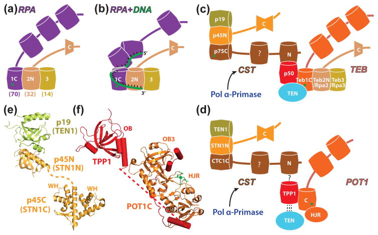
Telomerase is an RNP that synthesizes the 3' ends of linear chromosomes and is an important regulator of telomere length. It contains a single long non-coding telomerase RNA (TER), telomerase reverse transcriptase (TERT), and other proteins that vary among organisms. Recent progress in structural biology of telomerase includes reports of the first cryo-electron microscopy structure of telomerase, from Tetrahymena, new crystal structures of TERT domains, telomerase RNA structures and models, and identification in Tetrahymena telomerase holoenzyme of human homologues of telomere-associated proteins that have provided a more unified view of telomerase interaction at telomeres as well as insights into the role of telomerase RNA in activity and assembly.


Figures

Figure 1
Figure 1
Tetrahymena and human telomerase holoenzymes. (a) Front (left) and back (right) views of Tetrahymena telomerase cryo-EM map at ~9Å resolution (RNP core, blue; CST, tan; TEB, straw; and p50, red) and pseudoatomic models of the RNP core and TEB and CST trimerization domains of 3 OB-folds [13]. Tetrahymena telomerase protein domains for which structures have been determined are rendered as ribbons and with bases, respectively; homology models are rendered with ribbons and cylinders. TER single-stranded regions are shown as ribbons except the template which includes bases; folded domains are shown with bases. (b, d) Schematics illustrating (b) Tetrahymena and (d) human TER secondary structure and domains, TER interacting proteins, and proteins that interact with the RNP core. Proteins or complexes with generally homologous functions between organisms are colored the same, except H/ACA scaRNP proteins which are distinguished from each other. (c, e) Schematics of (c) Tetrahymena and (e) human interactions at telomere 3′ ends. Dashed green line indicates extension of the 3′ end by telomerase. In (e), lines with arrowheads indicate transient interactions and lines with bars indicate inhibitory interactions. Panels b-e are modified from [8].
Figure 2
Figure 2
Structure of TERT and interaction with TER. (a) Structure of flour beetle TERT with bound inhibitor BIBR1532 (PDB ID: 5CQG). The model is shown in the classical polymerase hand view (pale green) with RT palm (purple), fingers (orange), IFD (gold), CTE thumb (light blue), and TRBD (blue). Active site on RT is space-filled red. (b) Hand view of structure of Tetrahymena TERT with bound TER. Note that this view is rotated 180° at the Y-axis from the cryo-EM model back view in Figure 1a. (c) Tetrahymena TRBD-TBE complex (PDB ID: 5C9H), TBE is red, T motif is cyan, CP is gold, and CP2 is purple. (d) Human CTE (thumb) (PDB ID: 5UGW). (e) Model of human TERT bound to human TER t/PK based on the model of the human t/PK and the positions of homologous domains of Tetrahymena TR in the cryo-EM map. The dash line indicates the proposed location of P6.1. Subdomains of TERT and TR are colored as in Figure 1, except human TER J2a/b is colored as green.
Figure 3
Figure 3
Models for steps in Tetrahymena telomere DNA synthesis and assembly of TER with TERT. (a) Schematic illustration of secondary structure of Tetrahymena TER bound to p65 (left) and to p65 and TERT (right). In the p65 bound TER, the pseudoknot is sequestered in a helix that prevents it from folding, but the TBE and loop 4 remain accessible for subsequent binding to TERT. (b) Schematic illustrating DNA exit pathway and interaction with Teb1, based on [13]. (c) Schematics illustrating template movement through the active site at beginning and end of synthesis of a telomere repeat. The template boundary element interacts with TERT to physically restrain the RNA so that residues beyond the template are not copied (right). A proposed sstDNA retention surface (SRS) within the CTE is colored as dark blue [56]. (d) Schematic illustrating the DNA hairpin proposed to form after translocation as part of the telomere repeat synthesis cycle [55]. This model assumes a large movement of the CTE (thumb) domain during translocation, indicated by the arrowheads connected by dotted line. The “hands” in the panels indicate the orientation of TERT in the panels relative to the views in figure 2.
Figure 4
Figure 4
RPA, TEB, CST and their interactions. (a) Schematic of the RPA heterotrimer of Rpa1-Rpa2-Rpa3 (Rpa70, 32, and 14 in humans). Shared subunits Teb2/Rpa2 and Teb3/Rpa3 found in Tetrahymena are indicated by common colors with panel c. (b) Schematic of the structure of a RPA-ssDNA complex, based on [81]. (c) Schematic of the two RPA-like complexes, CST (p75-p45-p19) and TEB (Teb1-Teb2-Teb3), in Tetrahymena telomerase and their interactions with p50 and TEN domain; (d) Schematic of the two RPA-like complexes/proteins that transiently associate with human telomerase, CST (CTC1-Stn1-Ten1) and Pot1 (paralog of Rpa1), their interaction with TPP1 (putative for CTC1), and the interaction of TPP1 with the TEN domain. The human telomerase RNP core is recruited to telomere by TPP1-POT. CST interaction with TPP1-POT1 inhibits telomerase activity; (e,f) Crystal structures of (e) p45N-p19 complex (PDB ID: 5DOI) and p45C domain (PDB ID: 5DFN) and (f) POT1C-TPP1 complex which is modeled based on crystal structures of POT1C-TPP1 PBM complex (PDB ID: 5UN7), TPP1 (PDB ID: 2I46), and TEBPα–β–ssDNA complex (PDB ID: 1OTC) [65]. In (a-d), barrels indicate OB-fold domains, single trapezoid is WH domain, double trapezoid is tandem WH-WH domains, green star is a zinc-binding motif. Homologous domains for Tetrahymena and human telomerase/telomere proteins are colored the same (c-d); for RPA (a, b) the two smaller subunits are identical with those of TEB.

References

    1. Blackburn EH, Collins K. Telomerase: an RNP enzyme synthesizes DNA. Cold Spring Harb Perspect Biol. 2011;3:a003558. - PMC - PubMed
    1. Chen LY, Redon S, Lingner J. The human CST complex is a terminator of telomerase activity. Nature. 2012;488:540–544. - PubMed
    1. Wu RA, Upton HE, Vogan JM, Collins K. Telomerase Mechanism of Telomere Synthesis. Annu Rev Biochem. 2017;86:4.1–4.22. - PMC - PubMed
    1. Armanios M, Blackburn EH. The telomere syndromes. Nat Rev Genet. 2012;13:693–704. - PMC - PubMed
    1. Shay JW. Role of Telomeres and Telomerase in Aging and Cancer. Cancer Discov. 2016;6:584–593. - PMC - PubMed