. 2017 Jul 21;14(1):141.
doi: 10.1186/s12974-017-0920-8.

Sexually divergent induction of microglial-associated neuroinflammation with hippocampal aging

Affiliations

Sexually divergent induction of microglial-associated neuroinflammation with hippocampal aging

Colleen A Mangold et al. J Neuroinflammation. .

Abstract

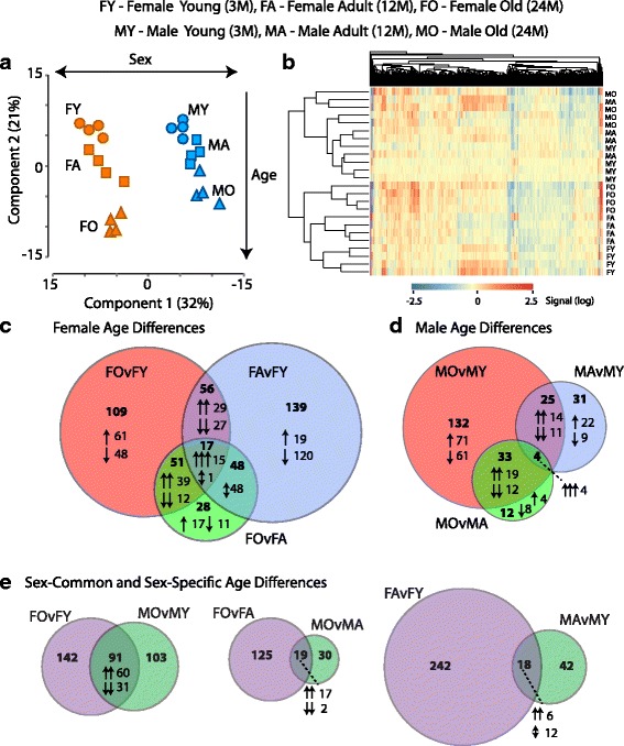
Background: The necessity of including both males and females in molecular neuroscience research is now well understood. However, there is relatively limited basic biological data on brain sex differences across the lifespan despite the differences in age-related neurological dysfunction and disease between males and females.

Methods: Whole genome gene expression of young (3 months), adult (12 months), and old (24 months) male and female C57BL6 mice hippocampus was analyzed. Subsequent bioinformatic analyses and confirmations of age-related changes and sex differences in hippocampal gene and protein expression were performed.

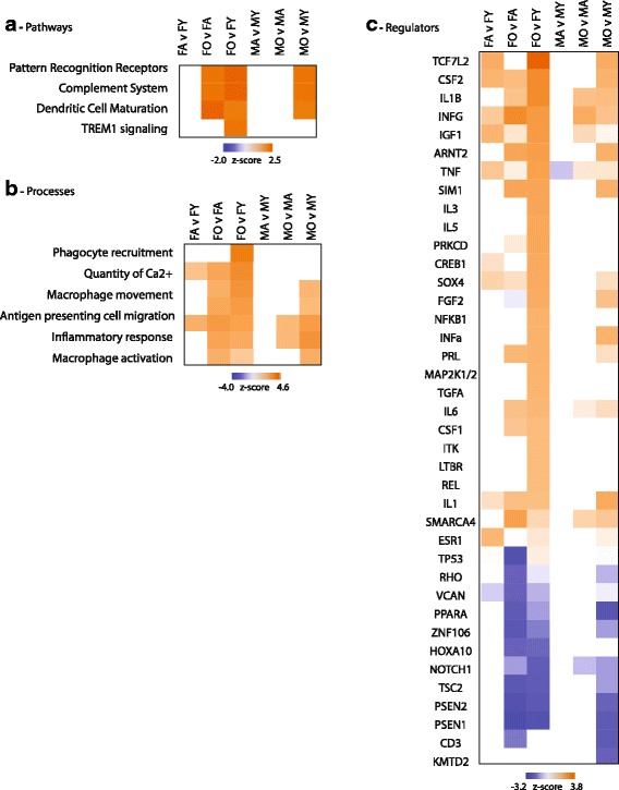
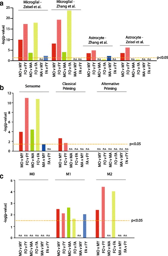
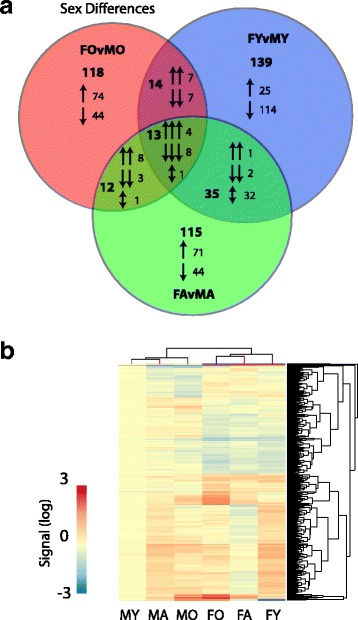
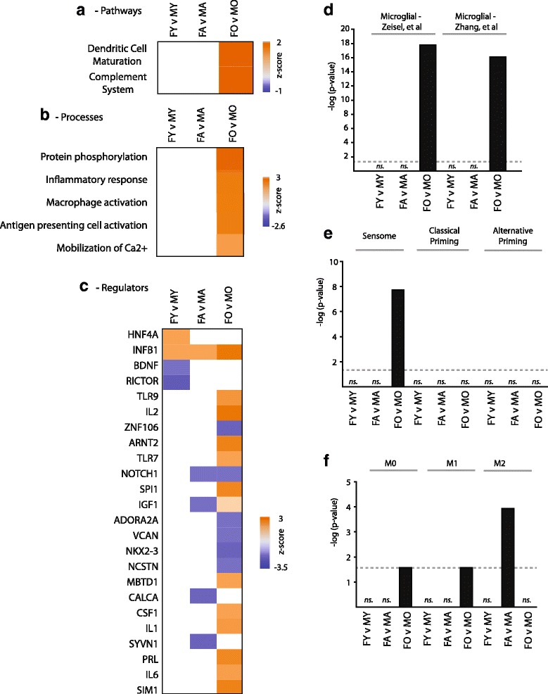
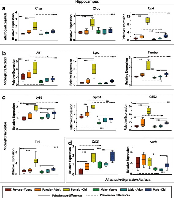
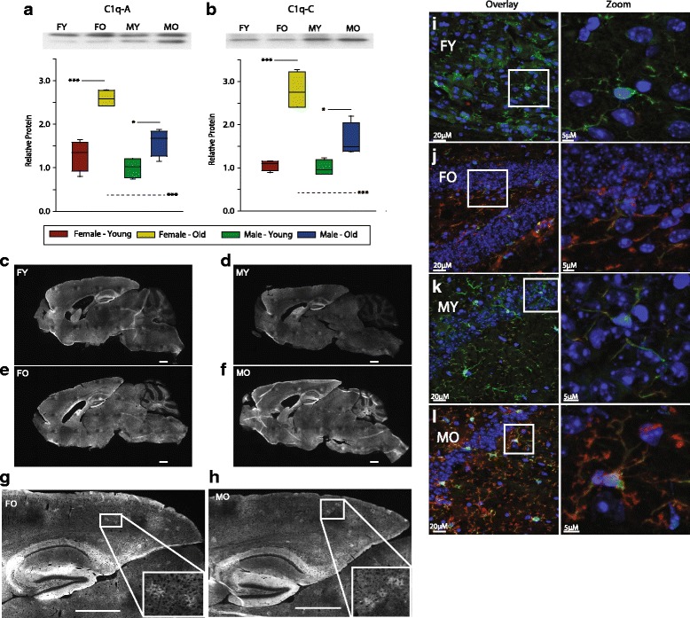
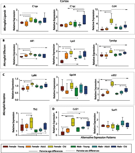
Results: Males and females demonstrate both common expression changes with aging and marked sex differences in the nature and magnitude of the aging responses. Age-related hippocampal induction of neuroinflammatory gene expression was sexually divergent and enriched for microglia-specific genes such as complement pathway components. Sexually divergent C1q protein expression was confirmed by immunoblotting and immunohistochemistry. Similar patterns of cortical sexually divergent gene expression were also evident. Additionally, inter-animal gene expression variability increased with aging in males, but not females.

Conclusions: These findings demonstrate sexually divergent neuroinflammation with aging that may contribute to sex differences in age-related neurological diseases such as stroke and Alzheimer's, specifically in the complement system. The increased expression variability in males suggests a loss of fidelity in gene expression regulation with aging. These findings reveal a central role of sex in the transcriptomic response of the hippocampus to aging that warrants further, in depth, investigations.

Keywords: Aging; Brain; Gene expression; Neuroinflammation; Sex differences.


Conflict of interest statement

Consent for publication

Not applicable

Competing interests

The authors declare that they have no competing interests.

Publisher’s Note

Springer Nature remains neutral with regard to jurisdictional claims in published maps and institutional affiliations.

Figures

Fig. 1
Fig. 1
Hippocampal gene expression changes with aging. a Principle component analysis (PCA) of individual samples using all expressed genes. Groups separated by sex along the 1st component and by age in the 2nd component. b Heatmap presentation of all age-related gene expression differences. Samples were clustered (Euclidean distance) by sex and age. c Comparison of pairwise age-related changes in females. Total numbers of genes and direction of change (induction/reduction). For intersections patterns in respective groups are also noted. d Venn diagram of pairwise aging changes in males. e Comparison of age-related differences between males and females
Fig. 2
Fig. 2
Gene expression variance with aging. a The distribution of inter-animal variance across ages was compared for females and males. Increasing variance with age was evident in males but not females. b Pairwise comparisons of variance between males and females at each age. Two-way ANOVA, SNK **p < 0.01, ***p < 0.001
Fig. 3
Fig. 3
Pathway, function, and regulatory analysis of age-related transcriptomic changes. Age-related gene expression changes were analyzed with Ingenuity Knowledge Base for differentially regulated pathways (a), functions (b), and regulators (c). A relevant selection of over-represented categories (Fisher’s exact test p < 0.05) is given in heatmap form with coloring according to the computed z score. Z scores are based on prior knowledge of known regulatory functions and direction of changes in the current dataset. Z scores >2 indicate significant activation with aging and <−2 indicate significant inhibition with aging. Abbreviations are detailed in Additional file 1: Table S8
Fig. 4
Fig. 4
Enrichment of age-related changes in cell-specific transcripts. a Cell-specific transcripts from previous reports (Zeisel et al. [30] and Zhang et al. [31]) were compared to each pairwise set of age-related changes. Fisher’s exact test p values are plotted for cell types with significant over-representations. b Using gene sets derived from Hickman et al. [32] for the sensome, classical priming, and alternative microglial priming, a significant over-representation of sensome genes, in particular, is evident. c Previously published gene sets indicative of M0, M1, and M2 microglial states [33] were also examined for over-representation of age-related genes
Fig. 5
Fig. 5
Hippocampal gene expression sex differences across the lifespan. a Sex-differences at each age are compared with the number of genes and direction of change (induction/reduction) noted. b Heatmap presentation of all sex differences in gene expression
Fig. 6
Fig. 6
Pathway, function, and regulatory analysis of sex differences in gene expression. A selection of statistically over-represented pathways (a), functions (b), and regulators (c) are presented with z scores is given in heatmap form with coloring according to the computed z score. Z scores are based on prior knowledge of known regulatory functions and direction of changes in the current dataset. Z scores >2 indicate significant activation in females as compared to males and <−2 indicate significant inhibition in females compared to males. d Cell-specific transcripts from previous reports [30, 31]) were compared to each pairwise set of sex differences. Fisher’s exact test p values are plotted for cell types with significant over-representations. Gen sets derived for the sensome, classical priming, and alternative microglial priming [32] (e),and gene sets indicative of M0, M1, and M2 microglial states (f) [33] were also examined for over-representation of age-related genes. Abbreviations are detailed in Additional File: Table S10
Fig. 7
Fig. 7
qPCR confirmation of differential sex- and age-related hippocampal gene expression. Selected microglial ligands (a), effectors (b), and receptors (c) targets identified in the microarray study were confirmed by gene-specific qPCR. Data is scaled to a mean value of 1 for young males. Boxes boundaries are the 25th and 75th percentiles, with median denoted by the bar and error bars at the 10th and 90th percentiles. Two-way ANOVA (age × sex), ***p < 0.001, **p < 0.01, *p < 0.05 Student–Newman–Kuels pairwise post hoc, n = 7–8/group. ANOVA values are presented in the text. Solid comparison lines denote age-related changes with a sex and dashed comparison lines are sex-related differences within an age. d A selection of genes with alternate expression parameters were also confirmed
Fig. 8
Fig. 8
Sexually divergent, age-related hippocampal C1q protein expression. Protein expression of compliment 1q isoforms C1qA (a) and C1qC (b) were induced with age and to a greater extent in females than males. Data is scaled to a mean value of 1 for young males. Boxes boundaries are the 25th and 75th percentiles, with median denoted by the bar and error bars at the 10th and 90th percentiles. Two-way ANOVA (age × sex), ***p < 0.001, *p < 0.05 Student–Newman–Kuels post hoc, n = 6/group. Solid comparison lines denote age-related changes with a sex, and dashed comparison lines are sex-related differences within an age. In sagittal brain sections qualitatively increased immunoreactivity for C1q was evident with aging across the brain in both female and male mice (cf). Detail regions of 24-month-old female (g) and male (h) mice show patches of C1qa-positive signals throughout the brain neuropil. Boxed area shows further magnified image to show the details of C1qa-positive patches. Scale bars 1 mm. C1q immunoreactivity was co-localized with Iba1 immunoreactivity in young females (i), old females (j), young males (k), and old males (l) demonstrating microglial expression. Scale bars 20 μm wide view, 5 μm zoomed view
Fig. 9
Fig. 9
Examine of sexually divergent gene expression in the cortex. To examine whether sexually divergent age-related changes in gene expression are evident across brain regions, selected microglial ligands (a), effectors (b), and receptors (c) targets from the hippocampus were examined in the cortex. Data is scaled to a mean value of 1 for young males. Boxes boundaries are the 25th and 75th percentiles, with median denoted by the bar and error bars at the 10th and 90th percentiles. Two-way ANOVA (age × sex), ***p < 0.001, **p < 0.01, *p < 0.05 Student–Newman–Kuels pairwise post hoc, n = 7–8/group. Solid comparison lines denote age-related changes within a sex, and dashed comparison lines are sex-related differences within an age. d A selection of genes with alternate expression parameters were also confirmed

References

    1. Kennedy BK, Berger SL, Brunet A, Campisi J, Cuervo AM, Epel ES, Franceschi C, Lithgow GJ, Morimoto RI, Pessin JE, et al. Geroscience: linking aging to chronic disease. Cell. 2014;159(4):709–713. doi: 10.1016/j.cell.2014.10.039. - DOI - PMC - PubMed
    1. Berchtold NC, Cribbs DH, Coleman PD, Rogers J, Head E, Kim R, Beach T, Miller C, Troncoso J, Trojanowski JQ, et al. Gene expression changes in the course of normal brain aging are sexually dimorphic. Proc Natl Acad Sci U S A. 2008;105(40):15605–15610. doi: 10.1073/pnas.0806883105. - DOI - PMC - PubMed
    1. Blalock EM, Grondin R, Chen KC, Thibault O, Thibault V, Pandya JD, Dowling A, Zhang Z, Sullivan P, Porter NM, et al. Aging-related gene expression in hippocampus proper compared with dentate gyrus is selectively associated with metabolic syndrome variables in rhesus monkeys. J Neurosci. 2010;30(17):6058–6071. doi: 10.1523/JNEUROSCI.3956-09.2010. - DOI - PMC - PubMed
    1. Kadish I, Thibault O, Blalock EM, Chen KC, Gant JC, Porter NM, Landfield PW. Hippocampal and cognitive aging across the lifespan: a bioenergetic shift precedes and increased cholesterol trafficking parallels memory impairment. J Neurosci. 2009;29(6):1805–1816. doi: 10.1523/JNEUROSCI.4599-08.2009. - DOI - PMC - PubMed
    1. Masser DR, Bixler GV, Brucklacher RM, Yan H, Giles CB, Wren JD, Sonntag WE, Freeman WM. Hippocampal subregions exhibit both distinct and shared transcriptomic responses to aging and nonneurodegenerative cognitive decline. J Gerontol A Biol Sci Med Sci. 2014;69(11):1311–1324. doi: 10.1093/gerona/glu091. - DOI - PMC - PubMed

Publication types

MeSH terms