

Diabetic retinopathy: Breaking the barrier
- PMID: 28732591
- PMCID: PMC5711541
- DOI: 10.1016/j.pathophys.2017.07.001
Diabetic retinopathy: Breaking the barrier
Abstract
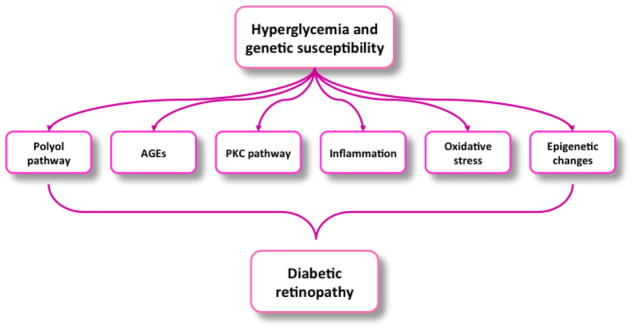
Diabetic retinopathy (DR) remains a major complication of diabetes and a leading cause of blindness among adults worldwide. DR is a progressive disease affecting both type I and type II diabetic patients at any stage of the disease, and targets the retinal microvasculature. DR results from multiple biochemical, molecular and pathophysiological changes to the retinal vasculature, which affect both microcirculatory functions and ultimately photoreceptor function. Several neural, endothelial, and support cell (e.g., pericyte) mechanisms are altered in a pathological fashion in the hyperglycemic environment during diabetes that can disturb important cell surface components in the vasculature producing the features of progressive DR pathophysiology. These include loss of the glycocalyx, blood-retinal barrier dysfunction, increased expression of inflammatory cell markers and adhesion of blood leukocytes and platelets. Included in this review is a discussion of modifications that occur at or near the surface of the retinal vascular endothelial cells, and the consequences of these alterations on the integrity of the retina.
Keywords: Blood-retinal barrier; Diabetes; Oxidative stress; Permeability; Retina.
Copyright © 2017 Elsevier B.V. All rights reserved.
Figures


References
-
- Retinal Conditions - Common Eye Disorders|Vision Health Initiative (VHI) 2014 cited; Available from: http://www.cdc.gov/visionhealth/basic_information/eye_disorders_retinal.htm.
-
- Fowler MJ. Microvascular and Macrovascular Complications of Diabetes. 2008 Apr 01;2008
-
- Morello CM. Etiology and natural history of diabetic retinopathy: an overview. Am J Health Syst Pharm. 2007 Sep 1;64(17 Suppl 12):S3–7. - PubMed
-
- Klein BE. Overview of epidemiologic studies of diabetic retinopathy. Ophthalmic Epidemiol. 2007 Jul-Aug;14(4):179–83. - PubMed
Publication types
Grants and funding
LinkOut - more resources
Full Text Sources
Other Literature Sources
