. 2017 Aug 23;37(34):8116-8130.
doi: 10.1523/JNEUROSCI.0908-17.2017. Epub 2017 Jul 21.

Pattern Analyses Reveal Separate Experience-Based Fear Memories in the Human Right Amygdala

Affiliations

Pattern Analyses Reveal Separate Experience-Based Fear Memories in the Human Right Amygdala

Senne Braem et al. J Neurosci. .

Abstract

Learning fear via the experience of contingencies between a conditioned stimulus (CS) and an aversive unconditioned stimulus (US) is often assumed to be fundamentally different from learning fear via instructions. An open question is whether fear-related brain areas respond differently to experienced CS-US contingencies than to merely instructed CS-US contingencies. Here, we contrasted two experimental conditions where subjects were instructed to expect the same CS-US contingencies while only one condition was characterized by prior experience with the CS-US contingency. Using multivoxel pattern analysis of fMRI data, we found CS-related neural activation patterns in the right amygdala (but not in other fear-related regions) that dissociated between whether a CS-US contingency had been instructed and experienced versus merely instructed. A second experiment further corroborated this finding by showing a category-independent neural response to instructed and experienced, but not merely instructed, CS presentations in the human right amygdala. Together, these findings are in line with previous studies showing that verbal fear instructions have a strong impact on both brain and behavior. However, even in the face of fear instructions, the human right amygdala still shows a separable neural pattern response to experience-based fear contingencies.SIGNIFICANCE STATEMENT In our study, we addressed a fundamental problem of the science of human fear learning and memory, namely whether fear learning via experience in humans relies on a neural pathway that can be separated from fear learning via verbal information. Using two new procedures and recent advances in the analysis of brain imaging data, we localized purely experience-based fear processing and memory in the right amygdala, thereby making a direct link between human and animal research.

Keywords: amygdala; fear; instructions; learning.


Figures

Figure 1.
Figure 1.
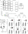
Procedure Experiment 1 and behavioral results. A, Experiment trials and rating screens. Each trial started with the presentation of a fixation cross, followed by the presentation of a CS. In the training phase, the CS+P was occasionally followed by a US (electrical stimulation) and the CS+U by a US placeholder (picture of a lightning bolt). The CS was never followed by either a US or placeholder. In the test phase, none of the CSs was followed by a US or placeholder. Rating screens assessed participant's fear experience and US expectancy associated with each of the CSs. B, Experimental procedure and instructions. Before training, subjects were instructed that only the CS+P could be followed by the US, while the CS+U could only be followed by the placeholder. Before testing, subjects were told to expect USs after both CS+s. Both phases consisted of three miniblocks (where every CS was randomly presented three times), each followed by a rating block. Three of nine CS+ presentations were followed by either the US or the US placeholder. C, Mean fear ratings. D, Mean US expectancy ratings. The error bars are ±1 SEM. tr, Training; te, test.
Figure 2.
Figure 2.
An example of the different types of similarity matrices in the anterior cingulate cortex (Experiment 1). A–C, The similarity matrices represent color-coded average Pearson correlation coefficients across subjects for every possible correlation between all different CS-presentation regressors in the trial-based model (A), miniblock model (B), or phase model (C). The vertical bars adjacent to each matrix indicate its color coding depending on the correlation coefficient.
Figure 3.
Figure 3.
Inter-CS similarities per region and experimental phase (Experiment 1), based on the miniblock model (Fig. 2). A–D, More similar multivoxel activation patterns between the two CS+s than between a CS+ and the CS indicate processing of learned stimulus value in the ACC (A), superior frontal gyrus (B; SFG), insula (C), and ventromedial prefrontal cortex (D; vmPFC). E, F, The amygdala does not exhibit greater similarity between the instructed and experienced CS+ (CS+P) and the merely instructed CS+ (CS+U) relative to the similarity between these CS+s and the CS. The error bars are ±1 SEM.
Figure 4.
Figure 4.
AF, Intra-CS similarities from miniblock to miniblock per CS type, region, and experimental phase (Experiment 1), based on the miniblock model (Fig. 2). Higher temporal consistency in intra-CS+ than intra-CS similarities indicates processing of learned stimulus qualities in a given ROI. This is not observed for the merely instructed CS+ (CS+U) in the amygdalae (E,F). The error bars are ±1 SEM.
Figure 5.
Figure 5.
A–F, Comparison of inter-CS similarities between each of the three different CS types from the test phase (CSte, CS+Pte, CS+Ute) and the CS+P pattern from the training phase (CS+Ptr), based on the phase model (Fig. 2), reveals CS+P-specific processing of threat-related information in the right amygdala (Experiment 1): CS+P-associated multivoxel activation patterns during test (CS+Pte) are more similar to CS+P-associated patterns during training (CS+Ptr) than CS+U-associated patterns during test (CS+Ute; F); other regions do not show such differentiation (A–E). The error bars are ±1 SEM. G, Individual differences in the difference between CS+PtrCS+Pte and CS+PtrCS+Ute inter-CS similarities in the right amygdala were correlated with differences in fear ratings between CS+P and CS+U in the test phase.
Figure 6.
Figure 6.
Inter-region similarity analyses between the ACC and left and right amygdalae (Experiment 1). A, Trial-by-trial intra-CS similarity matrices from the trial-based models for each region and CS type. On this basis, Spearman correlation coefficients were calculated between each combination of the resulting trial-by-trial ACC and left and right amygdala intra-CS similarity time courses, separately for each CS. B, C, The right amygdala showed a higher inter-region similarity with the ACC for the CS+P specifically (B), relative to the left amygdala, where no such effect was observed (C). The error bars are ±1 SEM.
Figure 7.
Figure 7.
Inter-region similarity analyses per different CS type and experimental phase. A, Correlations were calculated between each combination of two regions' intra-CS similarity matrices from the trial-based models (Fig. 2), per CS and phase separately, as explained in Figure 6. B, The correlations across all fear-related regions were averaged and are presented per CS type and phase separately. C, Visual two-dimensional scaling depiction of the similarities between different regions for each CS+ in the test phase separately.
Figure 8.
Figure 8.
Procedure Experiment 2 and results. A, Each trial consisted of a fear-contingency instruction and a CS presentation. The instruction indicated which of two pictures (CS+) would be followed by an electrical US by presenting an intensity meter next to that picture. A, lower left, Some CSs and instructions were recurring (old); others were always novel (new). Orthogonal to this, some instructions and subsequent CS presentations used pictures of houses, others of faces. CS+ presentation was always followed by a US presentation. On a small subset of trials, CS presentation was replaced by a catch question, to make certain that participants paid attention to the experiment. B, The similarity analyses focused exclusively on pattern similarities between face and house pictures during CS presentations, for each CS type separately (CS+old, CSold, CS+new, and CSnew). C–H, These analyses revealed that the right, but not the left, amygdala showed a differential response to fear relevance as a function of novelty. Namely, the pattern response for houses and faces were more similar when these denoted a CS+ than when they indicated a CS. However, this difference was only present when the CSs had been instructed and experienced before (old CSs), but disappeared when they were novel. The error bars are ±1 SEM.

References

    1. Atlas LY, Doll BB, Li J, Daw ND, Phelps EA (2016) Instructed knowledge shapes feedback-driven aversive learning in striatum and orbitofrontal cortex, but not the amygdala. Elife 5:e15192. 10.7554/elife.15192 - DOI - PMC - PubMed
    1. Bach DR, Weiskopf N, Dolan RJ (2011) A stable sparse fear memory trace in human amygdala. J Neurosci 31:9383–9389. 10.1523/JNEUROSCI.1524-11.2011 - DOI - PMC - PubMed
    1. Baker KB, Kim JJ (2004) Amygdalar lateralization in fear conditioning: evidence for greater involvement of the right amygdala. Behav Neurosci 118:15–23. 10.1037/0735-7044.118.1.15 - DOI - PubMed
    1. Bissière S, Plachta N, Hoyer D, McAllister KH, Olpe HR, Grace AA, Cryan JF (2008) The rostral anterior cingulate cortex modulates the efficiency of amygdala-dependent fear learning. Biol Psychiatry 63:821–831. 10.1016/j.biopsych.2007.10.022 - DOI - PMC - PubMed
    1. Carlson JM, Cha J, Mujica-Parodi LR (2013) Functional and structural amygdala–anterior cingulate connectivity correlates with attentional bias to masked fearful faces. Cortex 49:2595–2600. 10.1016/j.cortex.2013.07.008 - DOI - PubMed

LinkOut - more resources