

Immunohistochemical analysis of H3K27me3 demonstrates global reduction in group-A childhood posterior fossa ependymoma and is a powerful predictor of outcome
- PMID: 28733933
- PMCID: PMC5647236
- DOI: 10.1007/s00401-017-1752-4
Immunohistochemical analysis of H3K27me3 demonstrates global reduction in group-A childhood posterior fossa ependymoma and is a powerful predictor of outcome
Abstract
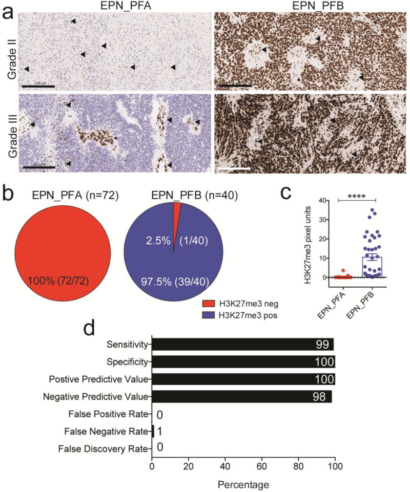
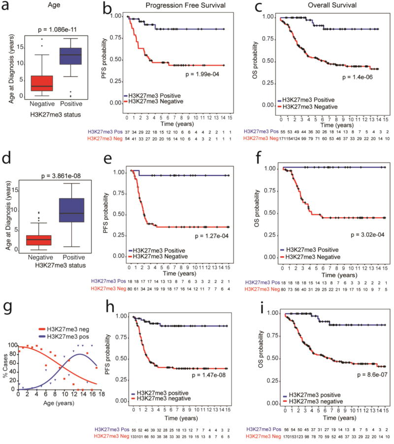
Posterior fossa ependymomas (EPN_PF) in children comprise two morphologically identical, but biologically distinct tumor entities. Group-A (EPN_PFA) tumors have a poor prognosis and require intensive therapy. In contrast, group-B tumors (EPN_PFB) exhibit excellent prognosis and the current consensus opinion recommends future clinical trials to test the possibility of treatment de-escalation in these patients. Therefore, distinguishing these two tumor subtypes is critical. EPN_PFA and EPN_PFB can be distinguished based on DNA methylation signatures, but these assays are not routinely available. We have previously shown that a subset of poorly prognostic childhood EPN_PF exhibits global reduction in H3K27me3. Therefore, we set out to determine whether a simple immunohistochemical assay for H3K27me3 could be used to segregate EPN_PFA from EPN_PFB tumors. We assembled a cohort of 230 childhood ependymomas and H3K27me3 immunohistochemistry was assessed as positive or negative in a blinded manner. H3K27me3 staining results were compared with DNA methylation-based subgroup information available in 112 samples [EPN_PFA (n = 72) and EPN_PFB tumors (n = 40)]. H3K27me3 staining was globally reduced in EPN_PFA tumors and immunohistochemistry showed 99% sensitivity and 100% specificity in segregating EPN_PFA from EPN_PFB tumors. Moreover, H3K27me3 immunostaining was sufficient to delineate patients with worse prognosis in two independent, non-overlapping cohorts (n = 133 and n = 97). In conclusion, immunohistochemical evaluation of H3K27me3 global reduction is an economic, easily available and readily adaptable method for defining high-risk EPN_PFA from low-risk posterior fossa EPN_PFB tumors to inform prognosis and to enable the design of future clinical trials.
Keywords: Childhood ependymoma; Epigenetics; H3K27me3; Molecular subgrouping.
Conflict of interest statement
Figures
Comment in
-
Neuro-oncology: A new approach to ependymoma subtyping.Nat Rev Neurol. 2017 Sep;13(9):512-513. doi: 10.1038/nrneurol.2017.114. Epub 2017 Aug 4. Nat Rev Neurol. 2017. PMID: 28776599 No abstract available.
References
-
- Bayliss J, Mukherjee P, Lu C, Jain SU, Chung C, Martinez D, Sabari B, Margol AS, Panwalkar P, Parolia A, et al. Lowered H3K27me3 and DNA hypomethylation define poorly prognostic pediatric posterior fossa ependymomas. Sci Transl Med. 2016;8:366ra161. doi: 10.1126/scitranslmed.aah6904. - DOI - PMC - PubMed
-
- Bechet D, Gielen GG, Korshunov A, Pfister SM, Rousso C, Faury D, Fiset PO, Benlimane N, Lewis PW, Lu C, et al. Specific detection of methionine 27 mutation in histone 3 variants (H3K27M) in fixed tissue from high-grade astrocytomas. Acta Neuropathol. 2014;128:733–741. doi: 10.1007/s00401-014-1337-4. - DOI - PMC - PubMed
-
- Bender S, Tang Y, Lindroth AM, Hovestadt V, Jones DT, Kool M, Zapatka M, Northcott PA, Sturm D, Wang W, et al. Reduced H3K27me3 and DNA hypomethylation are major drivers of gene expression in K27M mutant pediatric high-grade gliomas. Cancer Cell. 2013;24:660–672. doi: 10.1016/j.ccr.2013.10.006. - DOI - PubMed
Publication types
MeSH terms
Substances
Grants and funding
LinkOut - more resources
Full Text Sources
Other Literature Sources
Medical
