Review
. 2017 Aug;23(8):737-755.
doi: 10.1016/j.molmed.2017.06.004. Epub 2017 Jul 20.

Osteosarcoma: Molecular Pathogenesis and iPSC Modeling

Affiliations
Review

Osteosarcoma: Molecular Pathogenesis and iPSC Modeling

Yu-Hsuan Lin et al. Trends Mol Med. 2017 Aug.

Abstract

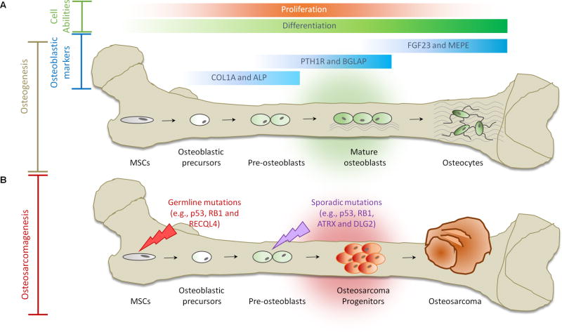
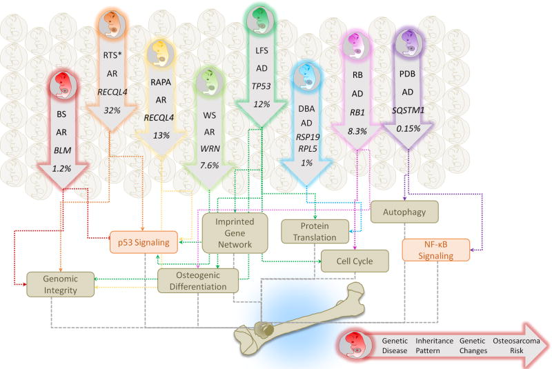
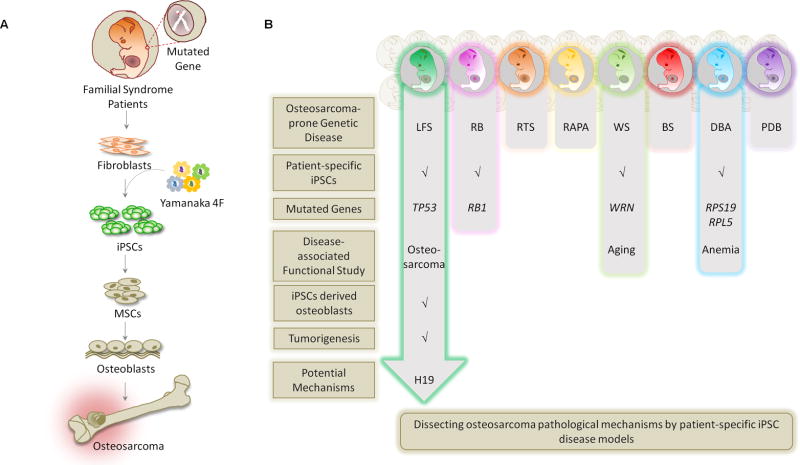
Rare hereditary disorders provide unequivocal evidence of the importance of genes in human disease pathogenesis. Familial syndromes that predispose to osteosarcomagenesis are invaluable in understanding the underlying genetics of this malignancy. Recently, patient-derived induced pluripotent stem cells (iPSCs) have been successfully utilized to model Li-Fraumeni syndrome (LFS)-associated bone malignancy, demonstrating that iPSCs can serve as an in vitro disease model to elucidate osteosarcoma etiology. We provide here an overview of osteosarcoma predisposition syndromes and review recently established iPSC disease models for these familial syndromes. Merging molecular information gathered from these models with the current knowledge of osteosarcoma biology will help us to gain a deeper understanding of the pathological mechanisms underlying osteosarcomagenesis and will potentially aid in the development of future patient therapies.

Keywords: cancer etiology; familial cancer syndrome; induced pluripotent stem cell; osteosarcoma.


Figures

Figure 1
Figure 1. Osteogenesis and Osteosarcomagenesis
(A) Initiation of osteogenic differentiation from MSCs. MSCs are multipotent bone marrow cells that are capable of differentiating to bone (osteoblast/osteocyte), fat (adipocyte) and cartilage (chondrocyte) tissues. Osteogenic differentiation is a tightly regulated process involving various signal transduction pathways (e.g., BMP and WNT), transcriptional regulators (e.g., p53, ZEB1, RUNX2 and ZNF521) and cell cycle controllers (e.g., RB1). Gene expression continuously changes through distinct osteogenic differentiation stages. COL1A and ALP are markers for osteoblastic progenitors and preosteoblasts. PTH1R and BGLAP serve as markers for mature osteoblasts. FGF23 and MEPE are markers for osteocytes. (B) Defects in osteogenesis lead to osteosarcomagenesis. Genetic alterations (e.g., germline mutations in p53, RB1 and RECQL4) probably interfere with the normal osteogenic process, resulting in incompletely differentiated osteoblasts or osteocytes in bone. These defects disrupt the balance between proliferation and differentiation and may cause a group of cells to display uncontrolled cell proliferation. Osteosarcoma progenitors may arise from these cells and expand to form osteosarcoma.
Figure 2
Figure 2. Familial Syndromes and Osteosarcoma
A cluster of familial syndromes predispose patients to the development of osteosarcomas and are of relevance to understanding the underlying genetics of these tumors. These include LFS, RB, RTS, RAPA, WS, BS, DBA, and PDB. Each inherited syndrome harbors distinct gene mutations but shares a cancer predisposition to osteosarcoma. AD, autosomal dominant; AR, autosomal recessive. The dysregulation of variant biological processes (e.g., imprinted gene network, osteogenic differentiation, genomic integrity, protein translation, cell cycle and autophagy) and signaling (e.g., p53 and NF-κB) contributes to the syndrome-associated osteosarcomagenesis. The syndrome-associated risk of osteosarcoma is stated as percentage of patients with disorder developing osteosarcoma. *RTS indicates RTS Type II.
Figure 3
Figure 3. Patient-Derived iPSC Disease Models for Osteosarcoma
(A) Patient-derived iPSCs are used to model human familial cancer syndromes and reveal a role of the mutant gene(s) in disease development. To apply iPSC methodology to study genetic disease-associated bone malignancy, patient fibroblasts are biopsied from skin and then reprogrammed to iPSCs by the four "Yamanaka factors" (OCT4, SOX2, KLF4 and c-MYC). iPSCs are then differentiated into MSCs, and then further, to osteoblasts. These iPSC-derived osteoblasts can be examined for osteoblast differentiation defects and tumorigenic ability. Systematic comparison of the genome/transcriptome/interactome between mutant and wild-type osteoblasts can further elucidate pathological mechanisms. (B) Current progress of applying LFS, RB, RTS, RAPA, WS, BS, DBA and PDB patient-derived iPSCs to model disease etiology and dissect disease-associated osteosarcomas.

References

    1. Ottaviani G, Jaffe N. The epidemiology of osteosarcoma. Cancer Treat Res. 2009;152:3–13. - PubMed
    1. Chen X, et al. Recurrent somatic structural variations contribute to turnorigenesis in pediatric osteosarcoma. Cell Rep. 2014;7:104–112. - PMC - PubMed
    1. Kansara M, et al. Translational biology of osteosarcoma. Nat Rev Cancer. 2014;14:722–735. - PubMed
    1. Stephens PJ, et al. Massive genomic rearrangement acquired in a single catastrophic event during cancer development. Cell. 2011;144:27–40. - PMC - PubMed
    1. Lorenz S, et al. Unscrambling the genomic chaos of osteosarcoma reveals extensive transcript fusion, recurrent rearrangements and frequent novel TP53 aberrations. Oncotarget. 2016;7:5273–5288. - PMC - PubMed

Publication types

MeSH terms