. 2017 Sep:62:200-206.
doi: 10.1016/j.neuro.2017.07.028. Epub 2017 Jul 20.

Methylmercury augments Nrf2 activity by downregulation of the Src family kinase Fyn

Affiliations

Methylmercury augments Nrf2 activity by downregulation of the Src family kinase Fyn

Megan Culbreth et al. Neurotoxicology. 2017 Sep.

Abstract

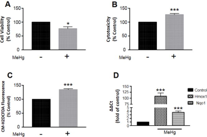
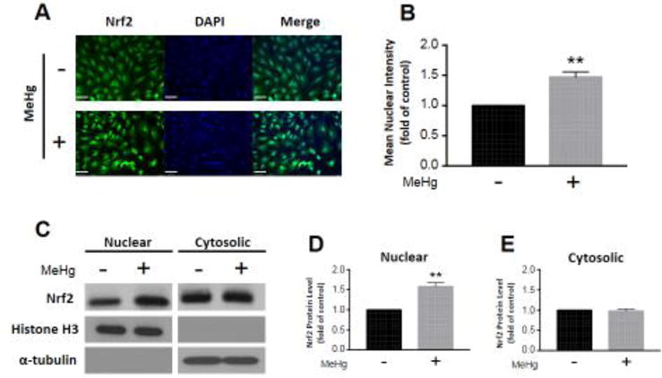
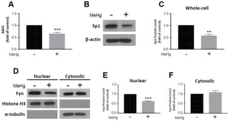
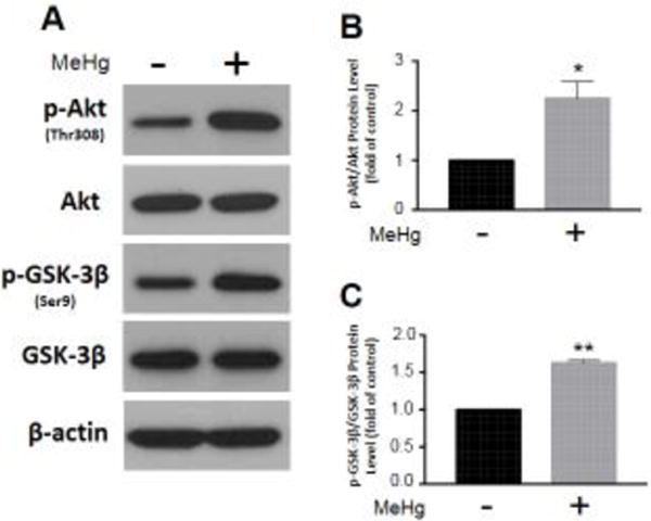
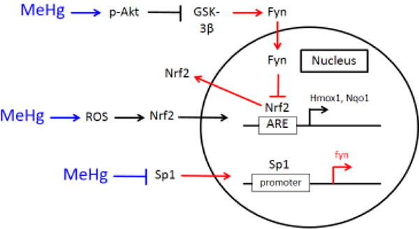
Methylmercury (MeHg) is a potent developmental neurotoxicant that induces an oxidative stress response in the brain. It has been demonstrated that MeHg exposure increases nuclear factor erythroid 2-related factor 2 (Nrf2) activity. Nrf2 is a transcription factor that translocates to the nucleus in response to oxidative stress, and upregulates phase II detoxifying enzymes. Although, Nrf2 activity is augmented subsequent to MeHg exposure, it has yet to be established whether Nrf2 moves into the nucleus as a result. Furthermore, the potential effect MeHg might have on the non-receptor tyrosine kinase, Fyn, has not been addressed. Fyn phosphorylates Nrf2 in the nucleus, resulting in its inactivation, and consequent downregulation of the oxidative stress response. Here, we observe Nrf2 translocates to the nucleus subsequent to MeHg-induced oxidative stress. This response is concomitant with reduced Fyn expression and nuclear localization. Moreover, we detected an increase in phosphorylated Akt and glycogen synthase kinase 3 beta (GSK-3β) at activating and inhibitory sites, respectively. Akt phosphorylates and inhibits GSK-3β, which subsequently prevents Fyn phosphorylation to signal nuclear import. Our results demonstrate MeHg downregulates Fyn to maintain Nrf2 activity, and further illuminate a potential mechanism by which MeHg elicits neurotoxicity.

Keywords: Fyn; Methylmercury; Nrf2; Oxidative stress.


Figures

Figure 1
Figure 1
MeHg-induced toxicity and oxidative stress are concomitant with Nrf2 activation of phase II detoxifying enzyme transcription. Astrocytes were treated with 5 μM MeHg for 6 hours, and cell viability (A; n =3) and cytotoxicity (B; n = 4) assessed by MTT and LDH assays, respectively. ROS generation (C; n = 4) was also measured by CM-H2DCFDA fluorescence, to indicate oxidative stress. Further, Nrf2 activation was confirmed by qRT-PCR of target genes Hmox1 (n = 5) and Nqo1 (n =6) (D). All data were normalized to untreated controls, and represent the mean ± SEM. *p < 0.05, ***p < 0.001
Figure 2
Figure 2
Nrf2 translocates to the nucleus in response to MeHg exposure. Astrocytes were treated with 5 μM MeHg for 6 hours, and immunostained for Nrf2 (A). Nrf2 mean intensity in cell nuclei was quantified from four distinct images (B; n = 4). Further, Nrf2 protein level was measured in nuclear (C,D; n = 3) and cytosolic fractions (C,E; n = 3) separated by subcellular fractionation. Representative blots for nuclear and cytosolic Nrf2, histone H3, and α-tubulin are displayed (C). Nuclear and cytosolic fractions were normalized to histone H3 and α-tubulin, respectively. All data were normalized to untreated controls, and represent the mean ± SEM. **p < 0.01, scale bar = 60 μm
Figure 3
Figure 3
Fyn expression and nuclear localization diminish consequent to MeHg exposure. Astrocytes were treated with 5 μM MeHg for 6 hours, and Fyn transcription (A; n = 6) assessed by qRT-PCR. Sp1 protein level was measured in whole-cell lysates, and normalized to β-actin (B,C; n = 3). Representative blots for Sp1 and β-actin are displayed (B). Further, Fyn protein level was quantified in nuclear (D,E; n = 3) and cytosolic fractions (D,F; n = 3) separated by subcellular fractionation. Representative blots for nuclear and cytosolic Fyn, histone H3, and α-tubulin are displayed (D). Nuclear and cytosolic fractions were normalized to histone H3 and α-tubulin, respectively. All data were normalized to untreated controls, and represent the mean ± SEM. **p < 0.01, ***p < 0.001
Figure 4
Figure 4
MeHg exposure alters the Akt/GSK-3β signaling pathway, which regulates Fyn nuclear import. Astrocytes were treated with 5 μM MeHg for 6 hours, and relevant protein levels assessed by whole-cell western blot. Representative blots for p-Akt(Thr308), Akt, p-GSK-3β(Ser9), GSK-3β, and β-actin are displayed (A). p-Akt (B; n = 3) and p-GSK-3β (C; n = 3) were normalized to total Akt and GSK-3β, respectively. All data were normalized to untreated controls, and represent the mean ± SEM. *p < 0.05, **p < 0.01
Figure 5
Figure 5
MeHg augments Nrf2 activation by downregulation of the Src family kinase Fyn. MeHg activates Akt, which in turn inhibits GSK-3β. Thus, GSK-3β does not signal Fyn nuclear import. ROS generation consequent to MeHg exposure promotes Nrf2 nuclear localization, which upregulates Hmox1 and Nqo1 transcription. Further, MeHg reduces Sp1 protein, which results in decreased Fyn gene expression. Blue arrows indicate the effects of MeHg, while red arrows designate processes disrupted subsequent to MeHg exposure.

References

    1. Abe J, Berk BC. Fyn and jak2 mediate ras activation by reactive oxygen species. J Biol Chem. 1999;274:21003–21010. - PubMed
    1. Alessi DR, James SR, Downes CP, Holmes AB, Gaffney PR, Reese CB, et al. Characterization of a 3-phosphoinositide-dependent protein kinase which phosphorylates and activates protein kinase balpha. Curr Biol. 1997;7:261–269. - PubMed
    1. Allen JW, Mutkus LA, Aschner M. Isolation of neonatal rat cortical astrocytes for primary cultures. Curr Protoc Toxicol Chapter. 2001;12 Unit12 14. - PubMed
    1. Bakir F, Damluji SF, Amin-Zaki L, Murtadha M, Khalidi A, al-Rawi NY, et al. Methylmercury poisoning in iraq. Science. 1973;181:230–241. - PubMed
    1. Charleston JS, Body RL, Mottet NK, Vahter ME, Burbacher TM. Autometallographic determination of inorganic mercury distribution in the cortex of the calcarine sulcus of the monkey macaca fascicularis following long-term subclinical exposure to methylmercury and mercuric chloride. Toxicol Appl Pharmacol. 1995;132:325–333. - PubMed

MeSH terms

LinkOut - more resources