. 2017 Aug 17;24(8):944-957.e7.
doi: 10.1016/j.chembiol.2017.06.009. Epub 2017 Jul 20.

β-TrCP1 Is a Vacillatory Regulator of Wnt Signaling

Affiliations

β-TrCP1 Is a Vacillatory Regulator of Wnt Signaling

Marcus John Long et al. Cell Chem Biol. .

Abstract

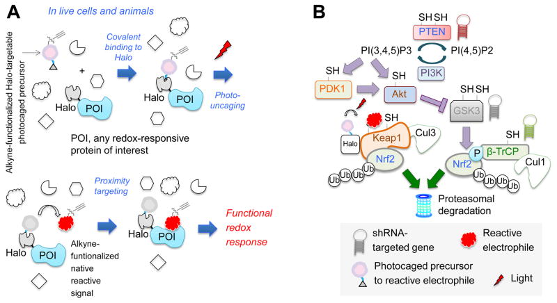
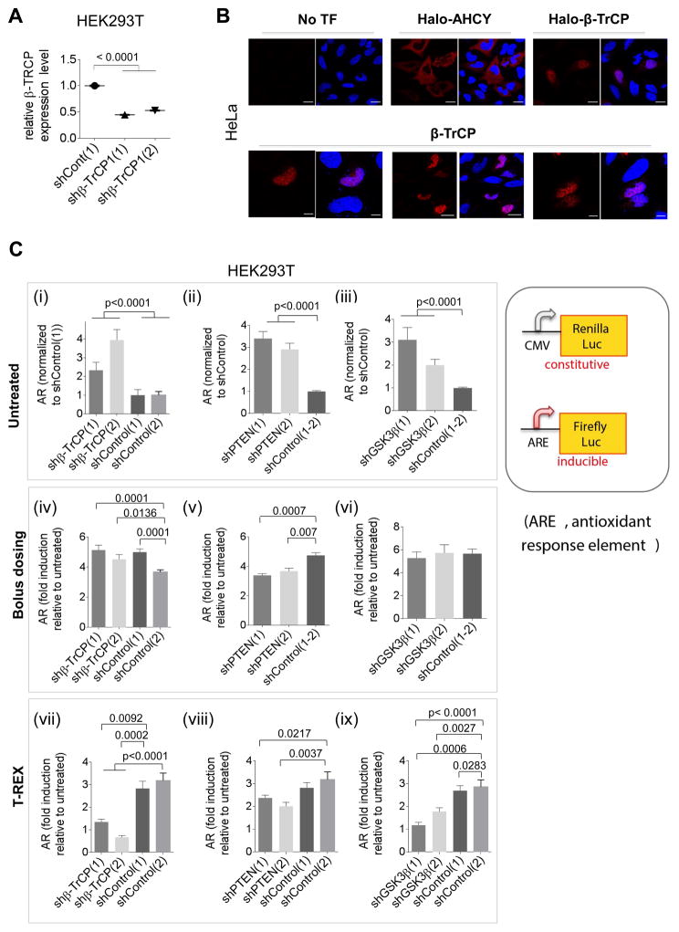
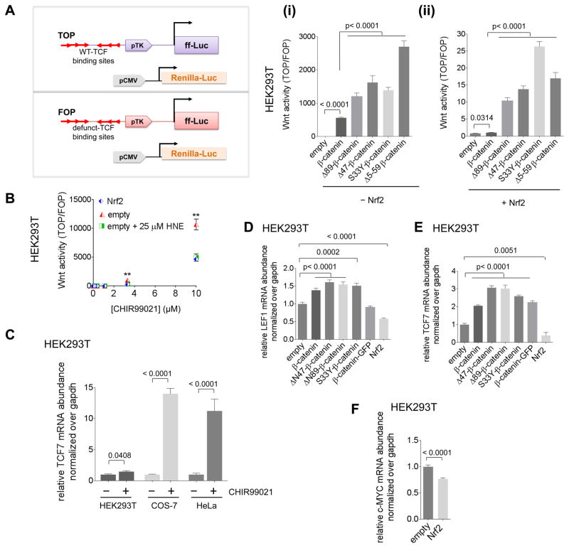
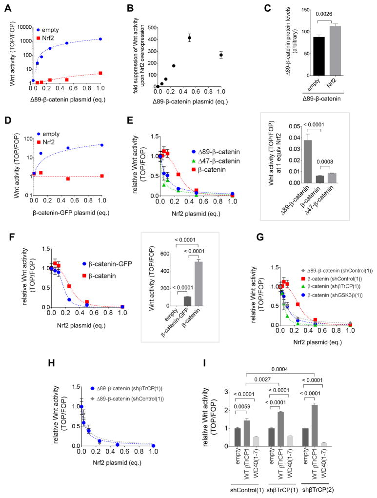
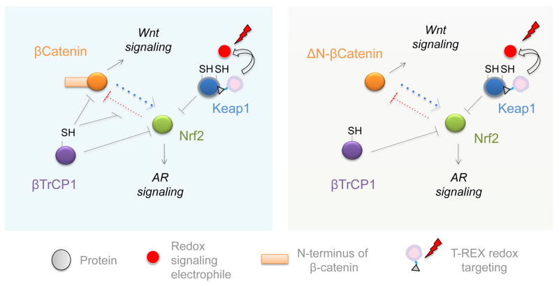
Simultaneous hyperactivation of Wnt and antioxidant response (AR) are often observed during oncogenesis. However, it remains unclear how the β-catenin-driven Wnt and the Nrf2-driven AR mutually regulate each other. The situation is compounded because many players in these two pathways are redox sensors, rendering bolus redox signal-dosing methods uninformative. Herein we examine the ramifications of single-protein target-specific AR upregulation in various knockdown lines. Our data document that Nrf2/AR strongly inhibits β-catenin/Wnt. The magnitude and mechanism of this negative regulation are dependent on the direct interaction between β-catenin N terminus and β-TrCP1 (an antagonist of both Nrf2 and β-catenin), and independent of binding between Nrf2 and β-TrCP1. Intriguingly, β-catenin positively regulates AR. Because AR is a negative regulator of Wnt regardless of β-catenin N terminus, this switch of function is likely sufficient to establish a new Wnt/AR equilibrium during tumorigenesis.

Keywords: 4-hydroxynonenal; HaloTag; Keap1-Nrf2-antioxidant response; reactive electrophile response; redox signaling; signaling crosstalk; β-TrCP; β-catenin/wnt signaling.


Figures

Figure 1
Figure 1. T-REX coupled with shRNA-knockdown investigates co-players in Keap1-alkylation-specific AR
(A) T-REX enables a user-controlled targeted redox perturbation in vivo: photocaged precursor to reactive LDE covalently binds HaloTag fused to any POI. After washing away unbound precursor, photouncaging rapidly (t1/2 < 1 min) releases LDE. Proximity enhancement (Long, et al., 2016) facilitates selective modification of first-responding sensor-POI, unveiling on-target redox responses. (B) Model of Nrf2 regulation. “SH”s mark redox-sensitive proteins. This study probes the functional impacts of T-REX- enabled Keap1-alklylation-specific redox events in cells in which either PTEN, GSK3β, or β-TrCP (marked by “hairpins”) is depleted.
Figure 2
Figure 2. Nuclear β-TrCP1 is necessary for Keap1-HNEylation-specific AR upregulation; bolus HNE exposure masks this requirement
(A) β-TrCP1 knockdown efficiency in two different shRNA-expressing lines from immunofluorescence (IF). Also see Figure S2. (B) Top panels: IF imaging in HeLa [anti-Halo (Table S1)] shows nuclear localization of Halo-β-TrCP1 as previously reported for β-TrCP1 (Seo, et al., 2009). AHCY—a known cytosolic protein—provides contrast (Hershfield, et al., 1985). Lower panels: Similar results were found with overexpression of untagged β-TrCP1 and IF imaging using β-TrCP antibody. Scale bar, 20 βm. (C) Effects of knockdowns, compared to non-targeted-shRNA controls, on the relative magnitude of Keap1-alkylation-specific AR. Note: for untreated [(i), (ii), (iii)], all signals are normalized to shControl; for other data [(iv)–(ix)], each bar is normalized to its respective value in the untreated set; the value in the untreated set is unity. Results from: untreated HEK293T cells [(i)–(iii); control cells set to 1]; bolus HNE dosing (25 βM HNE, 18 h) [(iv)–(vi); each relative to respective untreated set to 1]; T-REX (Keap1-specific-HNEyation) in cells [(vii)–(ix); each relative to respective untreated set to 1]. Inset: luciferase AR-reporters. The ratio of firefly (ARE) over Renilla (constitutive, CMV) gives AR. Data present Mean±s.d. with each bar graph from n>3 independent biological replicates.
Figure 3
Figure 3. β-catenin promotes AR regardless of its N-terminus
(A) AR fold-upregulation after T-REX-mediated Keap1-specific HNEylation in monoclonal HEK293T lines stably expressing a single copy of ARE::GFP that have been transfected with either Nrf2-wt or the three Nrf2–β-TrCP1-binding-defective mutants (Figure S3H), separately. See Figure S3J. (B) Nrf2-AR axis with a postulated novel modulator of Nrf2 that is regulated by β-TrCP1. (C) β-catenin stimulates AR regardless of its N-terminus. Post transfection with ARE::firefly luciferase, CMV::renilla, CMV::Nrf2 and the indicated plasmid in HEK293T, and AR was measured after 2 days. See also Figure S4B. (D) As in (C) except either wt or Δ89-β-catenin (balanced against an empty vector) was varied. (E) AR in HEK293T transfected with ARE::firefly luciferase, CMV::renilla, either empty plasmid or plasmid encoding ΔN89β-catenin, and varying amounts of CMV::Nrf2, measured after 2 days post transfection.
Figure 4
Figure 4. Dose response relationship between Wnt signaling and AR signaling
(A) Inset: Schematic of the TOP/FOP assay for Wnt activity: one set of cells is transfected with the TOP reporter, CMV::Renilla luciferase, empty vector [for 4A(i)], and indicated additional plasmid(s) as indicated; and a second set of cells is transfected with the FOP reporter, CMV::Renilla luciferase, Nrf2 plasmid [for 4A(ii)], and additional plasmid(s) as indicated. After 2 days, Wnt signaling was measured. Note: axes for 4A(i) and 4A(ii) are different. [The TOP construct contains 3 Wnt-specific binding sites for TCF (T-cell factor) transcription factors and the firefly (ff) luciferase (Luc) reporter under control of a minimal promoter. The FOP construct is identical to the TOP reporter but has the 3 TCF-binding sites mutated, rendering it inactive]. (B) At 1-day post-transfection of HEK293T with TOP or FOP, empty vector or Nrf2, and CMV::Renilla, samples were exposed to varying concentrations of CHIR99021 (with or without HNE) and after 24 h, Wnt activity was measured [Nrf2 (blue) and (empty vector + 25 μM HNE) (green) are coincidental for all concentrations of CHIR99021]. (C) The indicated cells were treated with either DMSO or CHIR99021 (10 μM) for 24 h, then TCF7 mRNA was measured using qRT-PCR. (D–F) HEK293T cells were transfected with plasmids encoding the indicated transgene. After 48 h, LEF1 (D), TCF7 (E), and C-MYC (F) mRNA were measured using qRT-PCR. Data present Mean ± s.e.m. with each bar graph from n>3 independent biological replicates.
Figure 5
Figure 5. Nrf2 is a negative regulator of Wnt signaling dependent on the β-catenin N-terminus
(A) HEK293T were transfected with TOP or FOP, CMV::Renilla, differing amounts of ΔN89-β-catenin (balanced with empty vector) with either empty vector or Nrf2. The y-axis is in log scale, making blue line appear curved but it best-fits a straight line (linear fit slope: 1500 ± 53, R2=0.995). (B) Data from (A) are shown as the ratio of Wnt signaling for empty vector/Nrf2 and expressed as a function of CMV::ΔN(89)-β-catenin dose. Wnt activity shows a hook/prozone effect upon titrating ΔN89-β-catenin in a Nrf2-overexpressing background. (C) HEK293T cells were transfected with ΔN(89)-β-catenin (Flag-tagged) together with either empty vector or Nrf2 (myc-tagged). After 48 h, levels of ΔN(89)-β-catenin were assessed by anti-Flag IF. (D) Same as (A) but β-catenin-GFP was used. The y-axis is in log scale, making blue line appear curved but it best-fits a straight line. (E) HEK293T cells expressing control shRNA were transfected with the indicated β-catenin construct, TOP or FOP, CMV::Renilla (see inset in Fig. 4A) and varying amounts of Nrf2 plasmid balanced with empty vector. After 48 h, Wnt signaling was assessed. Inset: At 1 equiv Nrf2, β-catenin N-terminal deletion mutants have higher Wnt activity than wt-β-catenin. (F) As in (E), except β-catenin with or without GFP fusion is compared over Nrf2 titration. Inset: HEK293T cells were transfected with TOP or FOP, the indicated β-catenin construct, and CMV::renilla, and Wnt signaling was measured after 48 h. (G) Same as (E) except indicated shRNA knockdown lines were used. Note: Nrf2 titration against wt-β-catenin in (E) is reshown in (F) and (I) for comparison. (H) Same as (E) except the indicated HEK293T lines were used. (I) The indicated HEK293T lines were transfected with wt-β-catenin, TOP or FOP, CMV::Renilla, 0.05 equivalents of Nrf2, and the plasmid encoding the indicated transgene or empty vector, and Wnt activity was measured after 48 h. (see β-TrCP-domain structure in Fig. S5I). Data present Mean ± s.e.m. with each bar graph from n>3 independent biological replicates.
Figure 6
Figure 6
β-TrCP1 regulates both β-catenin and Nrf2 stability through their GSK3β-regulated degrons. However, Nrf2 strongly inhibits β-catenin-dependent Wnt signaling, and β-catenin moderately stimulates Nrf2-dependent AR. The inhibition of β-catenin by Nrf2 is mediated by β-TrCP1-occupancy on β-catenin. Thus, cells with N-terminal mutated/truncated β-catenin that is unable to bind β-TrCP1 (ΔN-β-catenin) are much more susceptible to Nrf2/AR-mediated Wnt inhibition (Right Panel) than wt cells whose β-catenin contains a functional N-terminus (Left).

References

    1. Alomar SY, Mansour L, Abuderman A, Alkhuriji A, Arafah M, Alwasel S, Harrath AH, Almutairi M, Trayhyrn P, Dar JA. β-Catenin accumulation and S33F mutation of CTNNB1 gene in colorectal cancer in Saudi Arabia. Polish Journal of Pathology. 2016;2:156–162. - PubMed
    1. Blewett MM, Xie J, Zaro BW, Backus KM, Altman A, Teijaro JR, Cravatt BF. Chemical proteomic map of dimethyl fumarate-sensitive cysteines in primary human T cells. Sci Signal. 2016;9:rs10. - PMC - PubMed
    1. Bryan HK, Olayanju A, Goldring CE, Park BK. The Nrf2 cell defence pathway: Keap1-dependent and -independent mechanisms of regulation. Biochemical pharmacology. 2013;85:705–717. - PubMed
    1. Cadigan KM, Nusse R. Wnt signaling: a common theme in animal development. Genes & development. 1997;11:3286–3305. - PubMed
    1. Chan E, Gat U, McNiff JM, Fuchs E. A common human skin tumour is caused by activating mutations in [beta]-catenin. Nat Genet. 1999;21:410–413. - PubMed

MeSH terms