

Facile assembly and loading of theranostic polymersomes via multi-impingement flash nanoprecipitation
- PMID: 28736263
- PMCID: PMC5603398
- DOI: 10.1016/j.jconrel.2017.07.026
Facile assembly and loading of theranostic polymersomes via multi-impingement flash nanoprecipitation
Abstract
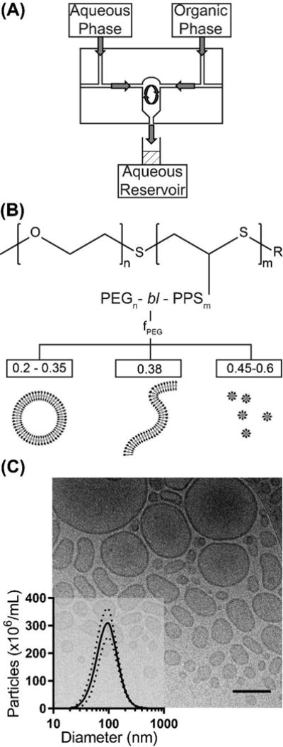
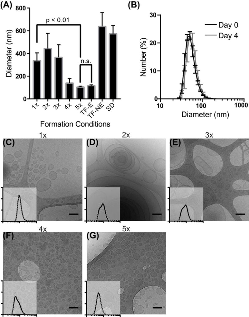
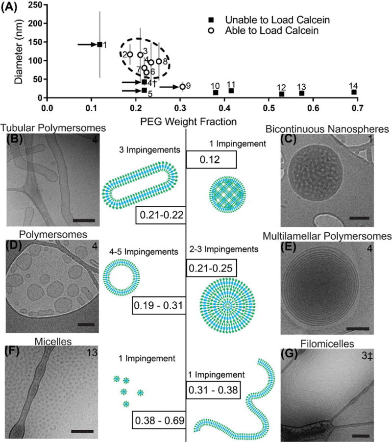
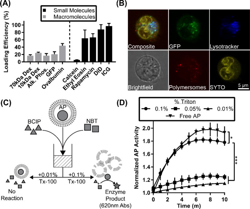
Flash nanoprecipitation (FNP) has proven to be a powerful tool for the rapid and scalable assembly of solid-core nanoparticles from block copolymers. The process can be performed using a simple confined impingement jets mixer and provides an efficient and reproducible method of loading micelles with hydrophobic drugs. To date, FNP has not been applied for the fabrication of complex or vesicular nanoarchitectures capable of encapsulating hydrophilic molecules or bioactive protein therapeutics. Here, we present FNP as a single customizable method for the assembly of bicontinuous nanospheres, filomicelles and vesicular, multilamellar and tubular polymersomes from poly(ethylene glycol)-bl-poly(propylene sulfide) block copolymers. Multiple impingements of polymersomes assembled via FNP were shown to decrease vesicle diameter and polydispersity, allowing gram-scale fabrication of monodisperse polymersomes within minutes. Furthermore, we demonstrate that FNP supports the simultaneous loading of both hydrophobic and hydrophilic molecules respectively into the polymersome membrane and aqueous lumen, and encapsulated enzymes were found to be released and remain active following vesicle lysis. As an example application, theranostic polymersomes were generated via FNP that were dual loaded with the immunosuppressant rapamycin and a fluorescent dye to link targeted immune cells with the elicited immunomodulation of T cells. By expanding the capabilities of FNP, we present a rapid, scalable and reproducible method of nanofabrication for a wide range of nanoarchitectures that are typically challenging to assemble and load with therapeutics for controlled delivery and theranostic strategies.
Keywords: Block copolymer; Drug delivery; Flash nanoprecipitation; Polymersome; Self-assembly.
Copyright © 2017 Elsevier B.V. All rights reserved.
Figures
Similar articles
-
Engineering endosomolytic nanocarriers of diverse morphologies using confined impingement jet mixing.Nanoscale. 2023 Oct 12;15(39):16016-16029. doi: 10.1039/d3nr02874g. Nanoscale. 2023. PMID: 37753868 Free PMC article.
-
Benchmarking Bicontinuous Nanospheres against Polymersomes for in Vivo Biodistribution and Dual Intracellular Delivery of Lipophilic and Water-Soluble Payloads.ACS Appl Mater Interfaces. 2018 Oct 10;10(40):33857-33866. doi: 10.1021/acsami.8b09906. Epub 2018 Sep 27. ACS Appl Mater Interfaces. 2018. PMID: 30213189
-
Flash nanoprecipitation permits versatile assembly and loading of polymeric bicontinuous cubic nanospheres.Nanoscale. 2018 Mar 15;10(11):5078-5088. doi: 10.1039/c7nr06779h. Nanoscale. 2018. PMID: 29255814 Free PMC article.
-
Application of flash nanoprecipitation to fabricate poorly water-soluble drug nanoparticles.Acta Pharm Sin B. 2019 Jan;9(1):4-18. doi: 10.1016/j.apsb.2018.11.001. Epub 2018 Nov 14. Acta Pharm Sin B. 2019. PMID: 30766774 Free PMC article. Review.
-
Biocompatible polymersomes-based cancer theranostics: Towards multifunctional nanomedicine.Int J Pharm. 2017 Mar 15;519(1-2):287-303. doi: 10.1016/j.ijpharm.2017.01.037. Epub 2017 Jan 20. Int J Pharm. 2017. PMID: 28115259 Review.
Cited by
-
Modulation of Schlemm's canal endothelial cell stiffness via latrunculin loaded block copolymer micelles.J Biomed Mater Res A. 2018 Jul;106(7):1771-1779. doi: 10.1002/jbm.a.36376. Epub 2018 Mar 6. J Biomed Mater Res A. 2018. PMID: 29468812 Free PMC article.
-
Surface engineered polymersomes for enhanced modulation of dendritic cells during cardiovascular immunotherapy.Adv Funct Mater. 2019 Oct 17;29(42):1904399. doi: 10.1002/adfm.201904399. Epub 2019 Aug 12. Adv Funct Mater. 2019. PMID: 34335131 Free PMC article.
-
Design of Modular, 3D-Printed Millifluidic Mixers to Enable Sequential NanoPrecipitation (SNaP) for the Tunable Synthesis of Drug-Loaded Nanoparticles and Microparticles.Adv Mater Technol. 2025 Jan 8;10(1):2400583. doi: 10.1002/admt.202570002. Epub 2024 Aug 3. Adv Mater Technol. 2025. PMID: 40814357 Free PMC article.
-
Engineering endosomolytic nanocarriers of diverse morphologies using confined impingement jet mixing.Nanoscale. 2023 Oct 12;15(39):16016-16029. doi: 10.1039/d3nr02874g. Nanoscale. 2023. PMID: 37753868 Free PMC article.
-
Induction of Mycobacterium Tuberculosis Lipid-Specific T Cell Responses by Pulmonary Delivery of Mycolic Acid-Loaded Polymeric Micellar Nanocarriers.Front Immunol. 2018 Nov 27;9:2709. doi: 10.3389/fimmu.2018.02709. eCollection 2018. Front Immunol. 2018. PMID: 30538700 Free PMC article.
References
-
- Duan X, Li Y. Physicochemical Characteristics of Nanoparticles Affect Circulation, Biodistribution, Cellular Internalization, and Trafficking. Small. 2013;9(9–10):1521–1532. - PubMed
-
- Stano A, Scott EA, Dane KY, Swartz MA, Hubbell JA. Tunable T cell immunity towards a protein antigen using polymersomes vs. solid-core nanoparticles. Biomaterials. 2013;34(17):4339–46. - PubMed
-
- Wang T, Bai J, Jiang X, Nienhaus GU. Cellular uptake of nanoparticles by membrane penetration: a study combining confocal microscopy with FTIR spectroelectrochemistry. ACS Nano. 2012;6(2):1251–9. - PubMed
MeSH terms
Substances
Grants and funding
LinkOut - more resources
Full Text Sources
Other Literature Sources
Research Materials
