

TFOS DEWS II Tear Film Report
- PMID: 28736338
- PMCID: PMC6035753
- DOI: 10.1016/j.jtos.2017.03.006
TFOS DEWS II Tear Film Report
Abstract
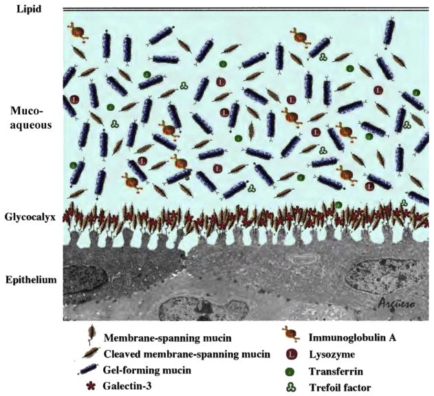
The members of the Tear Film Subcommittee reviewed the role of the tear film in dry eye disease (DED). The Subcommittee reviewed biophysical and biochemical aspects of tears and how these change in DED. Clinically, DED is characterized by loss of tear volume, more rapid breakup of the tear film and increased evaporation of tears from the ocular surface. The tear film is composed of many substances including lipids, proteins, mucins and electrolytes. All of these contribute to the integrity of the tear film but exactly how they interact is still an area of active research. Tear film osmolarity increases in DED. Changes to other components such as proteins and mucins can be used as biomarkers for DED. The Subcommittee recommended areas for future research to advance our understanding of the tear film and how this changes with DED. The final report was written after review by all Subcommittee members and the entire TFOS DEWS II membership.
Keywords: Dry eye disease; Evaporation; Lipidome; Mucin; Osmolarity; Proteome; Tear film; Tear film stability; Tears.
Copyright © 2017 Elsevier Inc. All rights reserved.
Figures
References
-
- Wolff E. The muco-cutaneous junction of the lid margin and the distribution of the tear fluid. Trans Ophthalmol Soc U K. 1946;66:291–308.
-
- Holly FJ, Lemp MA. Tear physiology and dry eyes. Surv Ophthalmol. 1977;22:69–87. - PubMed
-
- Doane MG. Abnormalities of the structure of the superficial lipid layer on the in vivo dry-eye. In: Sullivan DA, editor. Lacrimal Gland, Tear Film, and Dry Eye Syndromes. USA: Springer; 1994. pp. 489–93. - PubMed
-
- Yokoi N, Bron AJ, Georgiev GA. The precorneal tear film as a fluid shell: the effect of blinking and saccades on tear film distribution and dynamics. Ocul Surf. 2014;12:252–66. - PubMed
-
- Wang J, Aquavella J, Palakuru J, Chung S, Feng C. Relationships between central tear film thickness and tear menisci of the upper and lower eyelids. Invest Ophthalmol Vis Sci. 2006;47:4349–55. - PubMed
