Review
. 2017 Jul;15(3):404-437.
doi: 10.1016/j.jtos.2017.05.002. Epub 2017 Jul 20.

TFOS DEWS II pain and sensation report

Affiliations
Review

TFOS DEWS II pain and sensation report

Carlos Belmonte et al. Ocul Surf. 2017 Jul.

Abstract

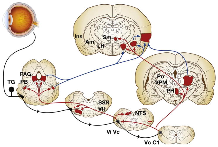
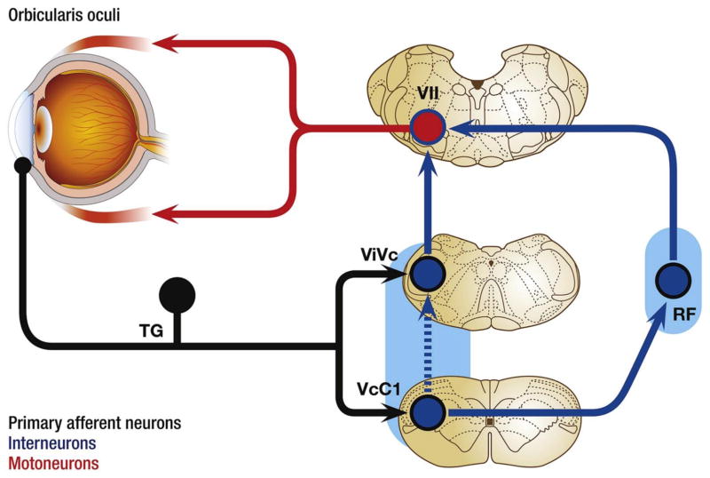
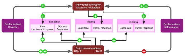
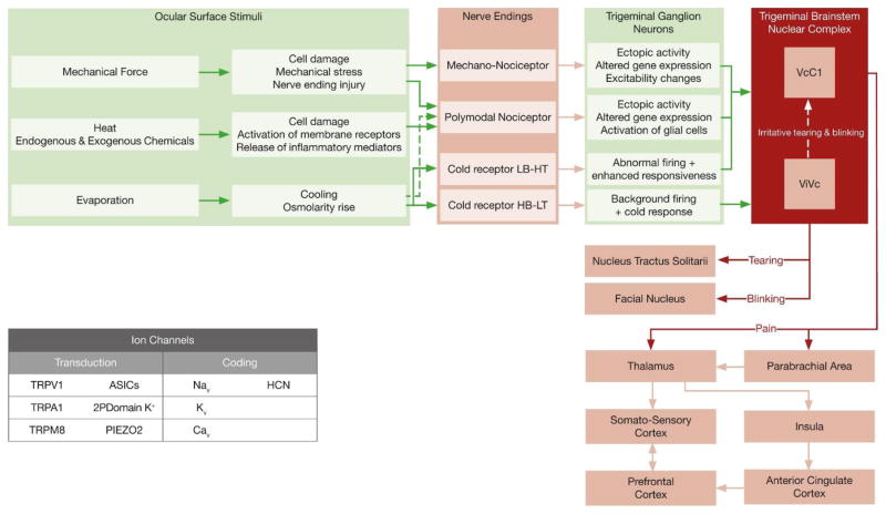
Pain associated with mechanical, chemical, and thermal heat stimulation of the ocular surface is mediated by trigeminal ganglion neurons, while cold thermoreceptors detect wetness and reflexly maintain basal tear production and blinking rate. These neurons project into two regions of the trigeminal brain stem nuclear complex: ViVc, activated by changes in the moisture of the ocular surface and VcC1, mediating sensory-discriminative aspects of ocular pain and reflex blinking. ViVc ocular neurons project to brain regions that control lacrimation and spontaneous blinking and to the sensory thalamus. Secretion of the main lacrimal gland is regulated dominantly by autonomic parasympathetic nerves, reflexly activated by eye surface sensory nerves. These also evoke goblet cell secretion through unidentified efferent fibers. Neural pathways involved in the regulation of meibomian gland secretion or mucin release have not been identified. In dry eye disease, reduced tear secretion leads to inflammation and peripheral nerve damage. Inflammation causes sensitization of polymodal and mechano-nociceptor nerve endings and an abnormal increase in cold thermoreceptor activity, altogether evoking dryness sensations and pain. Long-term inflammation and nerve injury alter gene expression of ion channels and receptors at terminals and cell bodies of trigeminal ganglion and brainstem neurons, changing their excitability, connectivity and impulse firing. Perpetuation of molecular, structural and functional disturbances in ocular sensory pathways ultimately leads to dysestesias and neuropathic pain referred to the eye surface. Pain can be assessed with a variety of questionaires while the status of corneal nerves is evaluated with esthesiometry and with in vivo confocal microscopy.

Keywords: Central nervous system; Cold receptors; Corneal esthesiometry; Dry eye disease; In vivo confocal microscopy; Mechano-nociceptors; Neuropathic pain; Ocular surface dryness; Pain; Peripheral sensory nerves; Polymodal nociceptors; Sensation; TRP channels; Trigeminal brainstem nuclear complex.


Figures

Fig. 1
Fig. 1
Medial view of the orbit, showing the sensory and autonomic innervation of the eye. The lacrimal gland has been removed for clarity. The ophthalmic branch (OB) of the trigeminal ganglion (TG) gives the nasociliary nerve (NCN) that sends long (LCN) and short (SCN) ciliary nerves to the eye ball. Frontal (FN) and lacrimal (LN) nerves appear as cut in this picture. Sympathetic fibers from the superior cervical ganglion, travelling in the carotid plexus (CPX) and parasympathetic branches of the ciliary (CG) and the pterigopalatine ganglion (PPG) join the short the ciliary nerves. OMN: Oculomotor nerve. MXN: Maxillary nerve. Modified from Netter, F, Atlas of Human Anatomy, 2nd Edition. Icon Learning Systems, 1997.
Fig. 2
Fig. 2
Reconstruction of superficial nerve terminals in the mouse corneal epithelium showing examples of simple (1,black), ramifying (2, red) and complex (3, blue) nerve terminals and impulse activity recorded from the different functional types of corneal nerve terminals in response to their specific stimuli (Modified from Ivanusic et al., [48] Gallar et al. [61] and Belmonte et al. [59,60]).
Fig. 3
Fig. 3
Major ascending brain pathways for trigeminal sensory fibers that supply the eye. The cell somata of sensory fibers are found within the trigeminal ganglion and project centrally to terminate in two spatially discrete regions of the trigeminal brainstem complex, the trigeminal subnucleus interpolaris/caudalis transition region (ViVc) and the caudalis/upper cervical cord junction (VcC1). Second-order ocular neurons in ViVc and VcC1 project to brain regions that mediate eyeblink (facial motor nucleus, VII), lacrimation (superior salivatory nucleus, SSN), and cardiovascular reflexes (nucleus tractus solitarius, NTS). Projections to higher centers such as the periaqueductal gray (PAG), PBA (PB), lateral hypothalamus (LH), posterior hypothalamus (PH), and amygdala (Am) contribute to the affective and modulatory aspects of ocular pain, while projections to posterior thalamus (posterior nuclear group, Po; ventral posteromedial nucleus, VPM) and insular cortex (Ins) mediate sensory-discriminative aspects. Note that a small group of ocular responsive neurons also are found in the contralateral ViVc; the source of input to this group is not well defined. Primary afferent fibers are drawn in black, second-order projections in red and third-order projections in blue. (Reproduced from Stapleton et al. [1]). (For interpretation of the references to colour in this figure legend, the reader is referred to the web version of this article.)
Fig. 4
Fig. 4
The corneal eye blink reflex is initiated by the free nerve endings in the cornea and involves the trigeminal nerve and ganglion (TG), the brainstem nuclei (VcC1: caudalis/upper cervical cord junction and ViVc: interpolaris/caudalis transition region), interneurons in the reticular formation (RF), motor neurons in the facial nucleus (VII) and nerve, and the orbicularis oculi. As the afferent information is distributed bilaterally to facial motor neurons by the reticular formation interneurons, the eye blink response is consensual, that is, both eye lids will close to stimulation of the cornea of either eye.
Fig. 5
Fig. 5
Schematic diagram summarizing how ocular inflammation of various etiologies or ocular surface dryness in DED, provoke variable increases (+) or decreases (−) of nerve impulse activity in polymodal- and mechano-nociceptors and in cold thermoreceptors of the high background, low threshold (HB-LT) and low background, high threshold (LB-HT) types. Together these changes evoke conscious sensations of different quality, as well as changes in tear flow and in spontaneous and reflex blinking.
Fig. 6
Fig. 6
Peripheral and central neural mechanisms involved in the sensory and autonomic responses evoked by eye surface dryness. The main types of ion channels involved in the transduction and coding of mechanical, thermal and chemical stimuli are represented in the inset.

References

    1. Stapleton F, Marfurt C, Golebiowski B, Rosenblatt M, Bereiter D, Begley C, et al. TFOS international Workshop on contact lens discomfort, the TFOS international Workshop on contact lens discomfort: report of the subcommittee on neurobiology. Investig Ophthalmol Vis Sci. 2013;54(11):TFOS71–TFOS97. - PMC - PubMed
    1. Belmonte C, Tervo TT. Pain in and around the eye. In: McMahon SB, Koltzenburg M, Tracey I, Turks DC, editors. Wall and Melzack’s Textbook of Pain. 6. Philadelphia, PA, USA: Elsevier Saunders; 2013.
    1. Wilson N. The semantics of pain in Greco-Roman antiquity. J Hist Neurosci. 2013;22(2):129–143. - PubMed
    1. IASP. The International Association for the Study of Pain
    1. von Hehn CA, Baron R, Woolf CJ. Deconstructing the neuropathic pain phenotype to reveal neural mechanisms. Neuron. 2012;73:638–652. - PMC - PubMed