Review
. 2017 Jul 7:11:192.
doi: 10.3389/fncel.2017.00192. eCollection 2017.

Resolution of Cochlear Inflammation: Novel Target for Preventing or Ameliorating Drug-, Noise- and Age-related Hearing Loss

Affiliations
Review

Resolution of Cochlear Inflammation: Novel Target for Preventing or Ameliorating Drug-, Noise- and Age-related Hearing Loss

Gilda M Kalinec et al. Front Cell Neurosci. .

Abstract

A significant number of studies support the idea that inflammatory responses are intimately associated with drug-, noise- and age-related hearing loss (DRHL, NRHL and ARHL). Consequently, several clinical strategies aimed at reducing auditory dysfunction by preventing inflammation are currently under intense scrutiny. Inflammation, however, is a normal adaptive response aimed at restoring tissue functionality and homeostasis after infection, tissue injury and even stress under sterile conditions, and suppressing it could have unintended negative consequences. Therefore, an appropriate approach to prevent or ameliorate DRHL, NRHL and ARHL should involve improving the resolution of the inflammatory process in the cochlea rather than inhibiting this phenomenon. The resolution of inflammation is not a passive response but rather an active, highly controlled and coordinated process. Inflammation by itself produces specialized pro-resolving mediators with critical functions, including essential fatty acid derivatives (lipoxins, resolvins, protectins and maresins), proteins and peptides such as annexin A1 and galectins, purines (adenosine), gaseous mediators (NO, H2S and CO), as well as neuromodulators like acetylcholine and netrin-1. In this review article, we describe recent advances in the understanding of the resolution phase of inflammation and highlight therapeutic strategies that might be useful in preventing inflammation-induced cochlear damage. In particular, we emphasize beneficial approaches that have been tested in pre-clinical models of inflammatory responses induced by recognized ototoxic drugs such as cisplatin and aminoglycoside antibiotics. Since these studies suggest that improving the resolution process could be useful for the prevention of inflammation-associated diseases in humans, we discuss the potential application of similar strategies to prevent or mitigate DRHL, NRHL and ARHL.

Keywords: age-related hearing loss; annexin A1; drug-induced hearing loss; galectin; inflammation; lipid mediators; noise-induced hearing loss; resolution of inflammation.


Figures

Figure 1
Figure 1
Potential fates for acute inflammation.
Figure 2
Figure 2
Temporal representation of the biochemical events associated with the onset and resolution of inflammation. The early phase of inflammation is characterized by the up-regulation (green arrows) of pro-inflammatory mediators such as leukotrienes (LTs), tumor necrosis factor-α (TNF-α), interleukin 1β (IL-1β), prostaglandin D2 (PGD2) and thromboxanes (TXs). Importantly, the anti-inflammatory mediator PGE2 is also up-regulated at this phase, indicating the controlled nature of inflammatory responses. The peak of the inflammatory response coincides with the start of the resolution phase, with down-regulation (blue arrows) of TXs, IL-1β, LTs and TNF-α, and up-regulation of anti-inflammatory cytokines such as PGE2, IL-10 and TGF-β; simultaneously, the synthesis and/or release of pro-resolution mediators (e.g., LX, resolvins, protectins, maresins, ANXA1) stop further infiltration of leukocytes and facilitate the removal of apoptotic cells, leading to the successful termination of the inflammatory response and the return to the tissue to its homeostatic condition. Modified from Maderna and Godson (2009).
Figure 3
Figure 3
Scheme showing the formation of lipoxins and aspirin-triggered lipoxins from arachidonic acid. The 2-D structures were obtained from the PubChem Substance and Compound database with the following chemical structure identifiers (CID): 444899 (AA), 5280914 (LXA4), 5280915 (LXB4), 9841438 (ATLXA4), 9928453 (ATLXB4; National Center for Biotechnology Information, 2017).
Figure 4
Figure 4
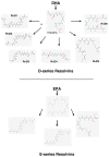
Scheme showing the biosynthesis of D-resolvins and E-resolvins from docosahexaenoic acid (DHA) and eicosapentaenoic acid (EPA), respectively. The 2-D structures were obtained from the PubChem Substance and Compound database with the following CID: 52921992 (17HpDHA), 16061135 (RvD1), 11383310 (RvD2), 53477497 (RvD3), 53477505 (RvD4), 16061139 (RvD5), 25073193 (RvD6), 446284 (18HpETE), 91820117 (RvE1), 16061125 (RvE2), 56848721 (RvE3; National Center for Biotechnology Information, 2017).
Figure 5
Figure 5
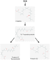
Scheme showing a simplified biosynthetic pathway for protectin 1 and protectin 2 from DHA. The 2-D structures were obtained from the PubChem Substance and Compound database with the following CID: 16061141 (16,17-epoxide-protectin), 16042541 (PRD1), 16061147 (PRD2; National Center for Biotechnology Information, 2017).
Figure 6
Figure 6
Scheme showing a simplified biosynthetic pathway for maresins. Macrophages convert DHA to the 13S-14S-epoxy-maresin intermediate, and from there MaR1, Mar2 and MaR3 are generated via soluble hydrolases. The 2-D structures were obtained from the PubChem Substance and Compound database with the following CID: 53477498 (14S-HpDHA), 60201795 (MaR1), 101894912 (MaR2), 52921996 (MaR3; National Center for Biotechnology Information, 2017).
Figure 7
Figure 7
Anti-inflammatory and pro-resolving effects of annexin A1 (ANXA1). Pharmacological administration of ANXA1 or its mimetic N-terminal peptides results in decreased migration and adhesion of leukocytes to the endothelium and inhibition of their passage through the walls of the blood vessels (diapedesis). In addition, ANXA1 is able to induce apoptosis, and clearance of apoptotic leukocytes by macrophages. Furthermore, ANXA1 promotes the reprogramming of macrophages toward a pro-resolving phenotype, and even the generation of non-professional phagocytic cells. Modified from Sugimoto et al. (2016b).
Figure 8
Figure 8
Diagram of a section of the cochlea. Note the three cochlear ducts, the scala vestibule (SV), the scala media (SM) and the scala tympani (ST). The auditory organ, the organ of Corti, sits at the SM. At the onset of inflammation, leukocytes and resident macrophages in the lateral wall and the spiral limbus migrate to the SV and the ST but, except in cases of extreme cochlear damage, they do not penetrate into the SM. Modified from Urrutia and Kalinec (2015).

References

    1. Abrashkin K. A., Izumikawa M., Miyazawa T., Wang C. H., Crumling M. A., Swiderski D. L., et al. . (2006). The fate of outer hair cells after acoustic or ototoxic insults. Hear. Res. 218, 20–29. 10.1016/j.heares.2006.04.001 - DOI - PubMed
    1. Aherne C. M., Collins C. B., Masterson J. C., Tizzano M., Boyle T. A., Westrich J. A., et al. . (2012). Neuronal guidance molecule netrin-1 attenuates inflammatory cell trafficking during acute experimental colitis. Gut 61, 695–705. 10.1136/gutjnl-2011-300012 - DOI - PMC - PubMed
    1. Aherne C. M., Kewley E. M., Eltzschig H. K. (2011). The resurgence of A2B adenosine receptor signaling. Biochim. Biophys. Acta 1808, 1329–1339. 10.1016/j.bbamem.2010.05.016 - DOI - PMC - PubMed
    1. Alessandri A. L., Sousa L. P., Lucas C. D., Rossi A. G., Pinho V., Teixeira M. M. (2013). Resolution of inflammation: mechanisms and opportunity for drug development. Pharmacol. Ther. 139, 189–212. 10.1016/j.pharmthera.2013.04.006 - DOI - PubMed
    1. Al-Gubory K. H., Fowler P. A., Garrel C. (2010). The roles of cellular reactive oxygen species, oxidative stress and antioxidants in pregnancy outcomes. Int. J. Biochem. Cell Biol. 42, 1634–1650. 10.1016/j.biocel.2010.06.001 - DOI - PubMed