Review
. 2017 Jun 22;2(6):281-292.
doi: 10.1302/2058-5241.2.160047. eCollection 2017 Jun.

Peroneal tendon disorders

Affiliations
Review

Peroneal tendon disorders

Kinner Davda et al. EFORT Open Rev. .

Abstract

Pathological abnormality of the peroneal tendons is an under-appreciated source of lateral hindfoot pain and dysfunction that can be difficult to distinguish from lateral ankle ligament injuries.Enclosed within the lateral compartment of the leg, the peroneal tendons are the primary evertors of the foot and function as lateral ankle stabilisers.Pathology of the tendons falls into three broad categories: tendinitis and tenosynovitis, tendon subluxation and dislocation, and tendon splits and tears. These can be associated with ankle instability, hindfoot deformity and anomalous anatomy such as a low lying peroneus brevis or peroneus quartus.A thorough clinical examination should include an assessment of foot type (cavus or planovalgus), palpation of the peronei in the retromalleolar groove on resisted ankle dorsiflexion and eversion as well as testing of lateral ankle ligaments.Imaging including radiographs, ultrasound and MRI will help determine the diagnosis. Treatment recommendations for these disorders are primarily based on case series and expert opinion.The aim of this review is to summarise the current understanding of the anatomy and diagnostic evaluation of the peroneal tendons, and to present both conservative and operative management options of peroneal tendon lesions. Cite this article: EFORT Open Rev 2017;2:281-292. DOI: 10.1302/2058-5241.2.160047.

Keywords: peroneal subluxation; peroneal tendinopathy; peroneal tendons; peroneus brevis; peroneus longus.


Conflict of interest statement

ICMJE Conflict of interest statement: P. O’Donnell reports that he receives royalties from a book published with Cambridge University Press.

Figures

Fig. 1
Fig. 1
Anatomy of the lateral ankle (reproduced with permission from Bentley G, ed. The European Surgical Orthopaedics and Traumatology The EFORT Textbook. Berlin: Springer Publications, 2014).
Fig. 2
Fig. 2
a-d) Sequential axial proton density (PD) MR images (a – d, superior to inferior). The peroneus brevis muscle (*) has a slender tendon (white arrow) that runs medial to the peroneus longus (black arrow) tendon in the distal calf (a) and maintain this relationship at the ankle (b). At the level of the talar dome (c), the superior peroneal retinaculum (white chevrons) attaches to the retromalleolar fibrocartilagenous ridge (white triangle) at the posterolateral aspect of the lateral malleolus. (d) Peroneus brevis tendon attaching to the base of the fifth metatarsal (white arrow).
Fig. 3
Fig. 3
a-b) Sagittal proton density MR images through the lateral aspect of the ankle, showing (a) the peroneus brevis (white arrows) and longus (black arrows) passing posterior to the lateral malleolus and the peroneus longus within the cuboid tunnel (black arrow, b).
Fig. 4
Fig. 4
a-b) Asymptomatic os peroneum. Axial (a) and sagittal (b) proton density MR images show a small focus of ossification (white triangle) in the peroneus longus tendon (white arrow) as it enters the cuboid tunnel. No oedema was visible in the ossicle on fluid sensitive sequences.
Fig. 5
Fig. 5
a-b) Oedematous os peroneum. Sagittal proton density fat suppression MR images lateral (a) and medial (b) showing an oedematous ossicle (white arrow) within the peroneus longus tendon (black arrow in (b)) and surrounding oedema fluid (*).
Fig. 6
Fig. 6
Axial proton density MR images demonstrating a peroneus quartus (black arrow) with either a distinct muscle belly (a) or slender tendon (b) posteromedial to peroneus brevis, with a separate and high variable distal insertion.
Fig. 7
Fig. 7
Anteroposterior radiograph demonstrating the ‘fleck’ sign (arrow) of the lateral malleolus due to acute peroneal tendon dislocation. Marked soft-tissue swelling is also seen here.
Fig. 8
Fig. 8
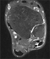
Axial proton density MR image with fat saturation demonstrating peroneal tenosynovitis. The peroneal brevis (B) and longus (L) appear swollen and there is excessive fluid in the tendon sheath (arrow).
Fig. 9
Fig. 9
Classification of peroneal tendon dislocation (reproduced with permission from Bentley G, ed. European Surgical Orthopaedics and Traumatology The EFORT Textbook. Berlin: Springer Publications, 2014).
Fig. 10
Fig. 10
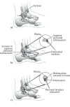
Surgical repair of acute peroneal tendon dislocation. a) Surgical approach. b) Pathological anatomy demonstrates dislocation of peroneal tendons. c) Repair of superior peroneal retinaculum (SPR) with drill holes through the posterolateral portion of fibula and reefing of SPR (reproduced with permission from Coughlin MJ, Schon LC. Disorders of tendons. In Coughlin MJ, Mann RA, Saltzman CL, eds. Mann’s Surgery of the Foot and Ankle. Ninth ed. Philadelphia: Mosby Elsevier, 2013:1264).
Fig. 11
Fig. 11
Operative exposure for groove-deepening procedure. Parallel saw cuts are used to create a trap door (b), which is hinged (c) posteriorly, exposing cancellous bone. d), a curette or burr is used to decompress this area. e) Trap door is impacted into place, creating an offset and deepening the recess posterior to the fibula. f) Peroneal tendons are relocated. g) Drill holes are placed in the fibular lip. h) Sutures are tied securing the superior peroneal retinaculum (reproduced with permission from Coughlin MJ, Schon LC. Disorders of tendons. In Coughlin MJ, Mann RA, Saltzman CL, eds. Mann’s Surgery of the Foot and Ankle. Ninth ed. Philadelphia: Mosby Elsevier, 2013:1273).
Fig. 12
Fig. 12
Intrasheath subluxation of the peroneal tendons. a) Normal position of peroneal brevis (1) and longus (2). b) Subluxation with peroneus longus deep to brevis. c) Peroneus brevis tear with subluxation of longus through the tear (reproduced with permission from Bentley G, ed. European Surgical Orthopaedics and Traumatology The EFORT Textbook. Berlin: Springer Publications, 2014).
Fig. 13
Fig. 13
Algorithm for the intra-operative assessment of peroneal tendon tear (reproduced with permission from Redfern D, Myerson M. The management of concomitant tears of the peroneus longus and brevis tendons. Foot Ankle Int 2004;25:695-707).

Similar articles

Cited by

References

    1. Dombek MF, Lamm BM, Saltrick K, Mendicino RW, Catanzariti AR. Peroneal tendon tears: a retrospective review. J Foot Ankle Surg 2003;42:250-258. - PubMed
    1. Maffulli N, Khan KM, Puddu G. Overuse tendon conditions: time to change a confusing terminology. Arthroscopy 1998;14:840-843. - PubMed
    1. Orthner E, Wagner M. Dislocation of the peroneal tendon. Sportverletz Sportschaden 1989;3:112-115. (In German) - PubMed
    1. Edwards M. The relations of the peroneal tendons to the fibula, calcaneus, and cuboideum. Am J Anat 1927;42:213-252.
    1. Mabit C, Salanne P, Boncoeur-Martel MP, et al. The lateral retromalleolar groove: a radio-anatomic study. Bull Assoc Anat (Nancy) 1996;80:17-21. (In French) - PubMed

LinkOut - more resources