

Detection theory for accurate and non-invasive skin cancer diagnosis using dynamic thermal imaging
- PMID: 28736673
- PMCID: PMC5516826
- DOI: 10.1364/BOE.8.002301
Detection theory for accurate and non-invasive skin cancer diagnosis using dynamic thermal imaging
Abstract
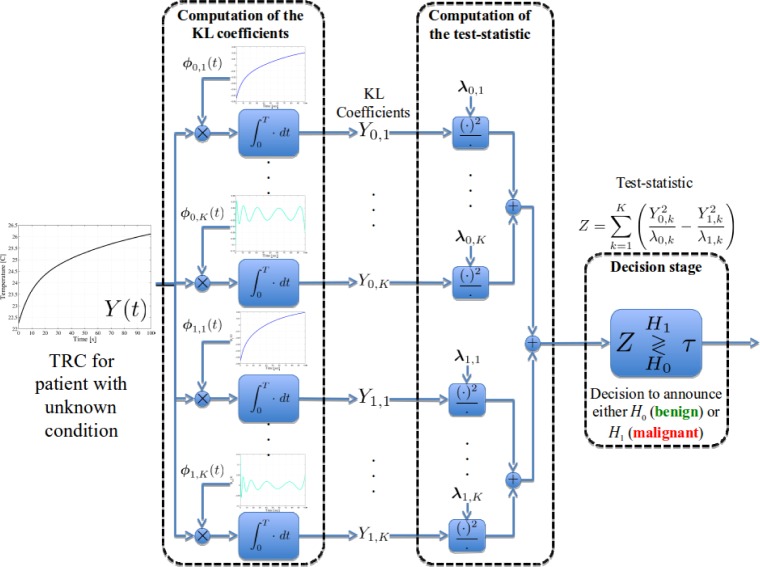
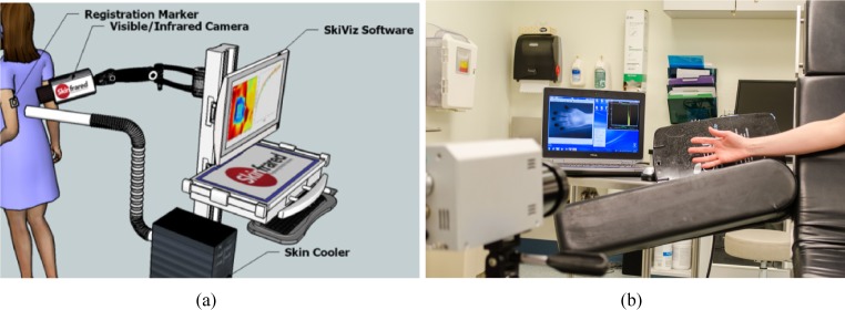
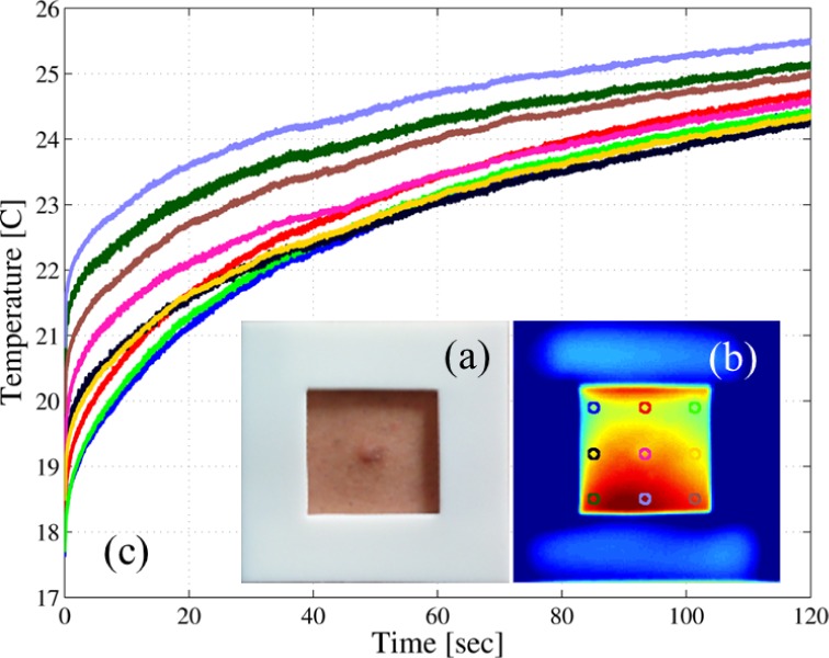
Skin cancer is the most common cancer in the United States with over 3.5M annual cases. Presently, visual inspection by a dermatologist has good sensitivity (> 90%) but poor specificity (< 10%), especially for melanoma, which leads to a high number of unnecessary biopsies. Here we use dynamic thermal imaging (DTI) to demonstrate a rapid, accurate and non-invasive imaging system for detection of skin cancer. In DTI, the lesion is cooled down and the thermal recovery is recorded using infrared imaging. The thermal recovery curves of the suspected lesions are then utilized in the context of continuous-time detection theory in order to define an optimal statistical decision rule such that the sensitivity of the algorithm is guaranteed to be at a maximum for every prescribed false-alarm probability. The proposed methodology was tested in a pilot study including 140 human subjects demonstrating a sensitivity in excess of 99% for a prescribed specificity in excess of 99% for detection of skin cancer. To the best of our knowledge, this is the highest reported accuracy for any non-invasive skin cancer diagnosis method.
Keywords: (040.1490) Cameras; (040.1880) Detection; (040.3060) Infrared; (110.2970) Image detection systems; (170.1610) Clinical applications; (170.3660) Light propagation in tissues; (170.3880) Medical and biological imaging; (170.4580) Optical diagnostics for medicine; (330.1880) Detection.
Figures
Similar articles
-
Role of In Vivo Reflectance Confocal Microscopy in the Analysis of Melanocytic Lesions.Acta Dermatovenerol Croat. 2018 Apr;26(1):64-67. Acta Dermatovenerol Croat. 2018. PMID: 29782304 Review.
-
Quantitative visualization and detection of skin cancer using dynamic thermal imaging.J Vis Exp. 2011 May 5;(51):2679. doi: 10.3791/2679. J Vis Exp. 2011. PMID: 21587160 Free PMC article.
-
The role of dynamic infrared imaging in melanoma diagnosis.Expert Rev Dermatol. 2013 Apr 1;8(2):177-184. doi: 10.1586/edm.13.15. Expert Rev Dermatol. 2013. PMID: 23745131 Free PMC article.
-
Screening for skin cancer.Am J Prev Med. 2001 Apr;20(3 Suppl):47-58. doi: 10.1016/s0749-3797(01)00258-6. Am J Prev Med. 2001. PMID: 11306232 Review.
-
Precision Diagnosis Of Melanoma And Other Skin Lesions From Digital Images.AMIA Jt Summits Transl Sci Proc. 2017 Jul 26;2017:220-226. eCollection 2017. AMIA Jt Summits Transl Sci Proc. 2017. PMID: 28815132 Free PMC article.
Cited by
-
An automatic approach to detect skin cancer utilizing active infrared thermography.Heliyon. 2024 Nov 26;10(23):e40608. doi: 10.1016/j.heliyon.2024.e40608. eCollection 2024 Dec 15. Heliyon. 2024. PMID: 39687094 Free PMC article.
-
Infrared thermal modulation endoscopy for label-free tumor detection.Sci Rep. 2024 Dec 30;14(1):31575. doi: 10.1038/s41598-024-76173-8. Sci Rep. 2024. PMID: 39738048 Free PMC article.
-
The Role of Artificial Intelligence in Early Diagnosis and Molecular Classification of Head and Neck Skin Cancers: A Multidisciplinary Approach.Diagnostics (Basel). 2024 Jul 10;14(14):1477. doi: 10.3390/diagnostics14141477. Diagnostics (Basel). 2024. PMID: 39061614 Free PMC article. Review.
-
A Method to Determine Human Skin Heat Capacity Using a Non-Invasive Calorimetric Sensor.Sensors (Basel). 2020 Jun 17;20(12):3431. doi: 10.3390/s20123431. Sensors (Basel). 2020. PMID: 32560551 Free PMC article.
-
Is thermography an effective screening tool for differentiating benign and malignant skin lesions in the head and neck? A systematic review.Arch Dermatol Res. 2024 Jun 15;316(7):404. doi: 10.1007/s00403-024-03166-y. Arch Dermatol Res. 2024. PMID: 38878184
References
-
- “American Cancer Society Cancer Facts & Figures 2014,” http://www.cancer.org/research/cancerfactsstatistics/cancerfactsfigures2.... Last Accessed: November, 2015.
-
- Benellii C., Roscetti E., Pozzo V. D., “The dermoscopic (7FFM) versus the clinical (ABCDE) diagnosis of small diameter melanoma,” Eur. J. Dermatol. 10, 282–287 (2000). - PubMed
LinkOut - more resources
Full Text Sources
Other Literature Sources