Review
. 2018 Jan;247(1):54-74.
doi: 10.1002/dvdy.24555. Epub 2017 Aug 30.

Diverse roles for glycosaminoglycans in neural patterning

Affiliations
Review

Diverse roles for glycosaminoglycans in neural patterning

Kristian Saied-Santiago et al. Dev Dyn. 2018 Jan.

Abstract

The nervous system coordinates the functions of most multicellular organisms and their response to the surrounding environment. Its development involves concerted cellular interactions, including migration, axon guidance, and synapse formation. These processes depend on the molecular constituents and structure of the extracellular matrices (ECM). An essential component of ECMs are proteoglycans, i.e., proteins containing unbranched glycan chains known as glycosaminoglycans (GAGs). A defining characteristic of GAGs is their enormous molecular diversity, created by extensive modifications of the glycans during their biosynthesis. GAGs are widely expressed, and their loss can lead to catastrophic neuronal defects. Despite their importance, we are just beginning to understand the function and mechanisms of GAGs in neuronal development. In this review, we discuss recent evidence suggesting GAGs have specific roles in neuronal patterning and synaptogenesis. We examine the function played by the complex modifications present on GAG glycans and their roles in regulating different aspects of neuronal patterning. Moreover, the review considers the function of proteoglycan core proteins in these processes, stressing their likely role as co-receptors of different signaling pathways in a redundant and context-dependent manner. We conclude by discussing challenges and future directions toward a better understanding of these fascinating molecules during neuronal development. Developmental Dynamics 247:54-74, 2018. © 2017 Wiley Periodicals, Inc.

Keywords: axon guidance; cell migration; chondroitin sulfate; code; dermatan sulfate; glycosaminoglycans; heparan sulfate; keratan sulfate; neuron; synapse.


Figures

Figure 1
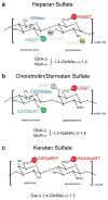
Figure 1. Disaccharide units and modifications of glycosaminoglycans (GAGs)
Schematics of heparan sulfate (HS)(A), chondroitin/dermatan sulfate (CS/DS)(B), and keratan sulfate (KS)(C) disaccharide repeats. HS modification enzymes and the specific positions they modify are shown as: NDSTs, (N-deacetylase-N-sulfotransferases); HS5epi, (C5-glucuronyl-epimerase); HS2ST, HS-2-O-sulfotransferase; HS3STs, HS-3-O-sulfotransferases; HS6STs, HS-6-O-sulfotransferases. CS/DS modification enzymes and the specific positions they modify are shown as: CS5epi, (CS-C5-epimerase); CS/DS2ST (UST), (CS/DS-2-O-sulfotransferase); CS/DS4STs, (CS/DS-4-O-sulfotransferases); CS/DS6STs, (CS/DS-6-O-sulfotransferases). KS modification enzymes and the specific positions they modify are shown as: GlcNAc6ST, (N-acetyl-glucosamine-6-O-sulfotransferase); KSGal6ST, (KS-galactose-6-O-sulfotransferase). Abbreviations used: GlcA, glucuronic acid; IdoA, iduronic acid; GlcNAc, N-acetyl-glucosamine; GalNAc, N-acetyl-galactosamine; Gal, galactose. Note that GAG chains often contain dozens of disaccharides, which are modified in a non-uniform manner, i.e. not all positions that can be modified, are modified.
Figure 2
Figure 2. HS/HSPGs serve specific roles in neuronal development in C. elegans
A. Schematics of the stereotypical axon extension of PVQL/R in wild-type and different HS mutant animals is shown (top). The red lines depict representative axon guidance defects in mutant animals. Note that all HSPG schematics here in and in other figures are for general information only. Actual number of GAG side chains and other structural characteristics may vary between species. A model displaying the genetic interaction between SLT-1/slit, SAX-3/Robo and HS/HSPGs in PVQ axon guidance is shown (bottom). B. Schematics of axon extensions of DA and DB neurons in wild-type animals and in animals with ectopic expression of the hst-6/6-O-sulfotransferase in the hypodermis (skin) (left). Green lines depict the correct axon extension of DA3, DB3 and DA4 neurons. Red lines depict the defects observed in animals with ectopic HS 6-O-sulfation. C. Schematics of the stereotypical HSN migration (left) and axon guidance (right) in wild-type (top) and different HS mutants (bottom). Green lines and arrows denote the normal and stereotypical migration or axon guidance of the HSN neurons in the body of the animal while the red lines and arrows denote migration or axon guidance defects.
Figure 3
Figure 3. Retinal axon guidance at the optic chiasm is coordinated by HS and CS glycans
A. Schematic demonstrating the stereotypical extension of ipsilateral (dark green) and contralateral axons (light green) across the optic chiasm in wild-type mice. B. Schematic demonstrating different misrouting defects of ipsilateral axons (orange) and contralateral axons (red) in animals lacking HS modifications or CS. Specific HS modifications guide contralateral retinal axons towards the optic tract. HS 2-O-sulfation is required to maintain axons organized in bundles and at the chiasm. HS 6-O-sulfation is required to prevent axons from extending away from the chiasm and the optic tract. Asterisks denote defects observed in a double mutant with Slit1. CS inhibits the crossing of ipsilateral axons at the midline in the optic chiasm and repels contralateral axons towards the optic tract. C. Schematic showing the expression of HS/CS glycans with important morphogens at the optic chiasm. The expression pattern of particular HS epitopes in and around the chiasm is highlighted: HS 2-O-sulfated moieties (indicated as green bubbles) are abundant throughout the optic chiasm, and high levels of this modification concentrate at regions where the optic nerve reaches the brain; HS 6-O-sulfated moieties (indicated as red bubbles) show a unique expression pattern at the chiasm, concentrating at the borders of this structure. Slit1 and Slit2 (colored hexagons) expression correlates with the HS 2-O-sulfated and HS 6-O-sulfated structures at the chiasm, respectively. The interaction between this network of genes guides retinal axons in a context-dependent manner. Similar to the expression pattern observed at the dorsal and ventral borders in HS 6-O-sulfated glycans, Shh (blue circles) is found off the chiasm. CS glycans (blue chains) are found at the midline of the chiasm, sharing a similar expression pattern to the EphB2 ligand (triangles). Both of these molecules are crucial for the turning of ipsilateral axons, hinting at a possible interaction between them.
Figure 4
Figure 4. Functions of HS and HSPGs in formation and function of the Drosophila NMJ
HS and HSPGs are located at the pre-synaptic, post-synaptic and synaptic cleft (sub-synaptic reticulum) of the Drosophila NMJ. Distinct HS proteoglycans and HS modifications have been shown to regulate the formation of boutons and, synaptic transmission at NMJs. (From left to right) Wnt-signaling is crucial for the formation of pre- and post-synaptic terminals. The secreted proteoglycan Trol/Perlecan regulates many of these Wnt-dependent functions by modulating the expression of Wg/Wnt in a HS-dependent manner. HS chains containing 6-O-sulfated epitopes also control the abundance of Wg/Wnt and gbb/BMP ligands at the synapse. The membrane-bound proteoglycans Sdc/Syndecan and Dlp/Glypican interact with the LAR receptor to promote synaptic and active zone formation, respectively. Furthermore, processing of Dlp/glypican by two distinct metalloproteinases modulates its expression and function at the NMJ. Synaptic transmission and homeostasis relies on a putative HSPG present at the synaptic cleft, Multiplexin/collagen XVIII. Different domains of Multiplexin/collagen XVIII modulate calcium channels at the pre-synaptic terminal. Abbreviations used: ndst, N-deacetylease-N-sulfotransferase; ext, exostosis; sulf1, HS 6-O-sulfatase; MMP, metalloproteinase.
Figure 5
Figure 5. Conserved and redundant HS mechanisms shape distinct aspects of neuronal development
A. Schematics in each panel illustrate the functional interactions between HS/HSPGs and their putative GAG chains with the Slit-Robo cassette in midline crossing and axonal pathfinding. Individual panels summarize the current knowledge regarding the interactions between these signaling networks in different model organisms. Studies in different model organisms suggest that the molecules and mechanisms highlighted here have been preserved during evolution to mediate the process of axon guidance. B. Schematics in each panel illustrate the functional interactions between HS/HSPGs with Wnt-signaling genes in different neuro-developmental processes. Recent findings propose that HS is a fundamental co-factor regulating these signaling pathways during neuronal development in a context-dependent manner. An example of this regulation is observed in interactions between HS/HSPGs and Wnt-signaling genes in neuronal migration, axon guidance and synaptic formation in distinct invertebrate model organisms. HS is also known to regulate the FGF and Slit/Robo pathways in an analogous fashion.

References

    1. Ackley BD, Crew JR, Elamaa H, Pihlajaniemi T, Kuo CJ, Kramer JM. The NC1/Endostatin Domain of Caenorhabditis elegans Type XVIII Collagen Affects Cell Migration and Axon Guidance. J Cell Biol. 2001;152:1219–1232. - PMC - PubMed
    1. Akita K, Toda M, Hosoki Y, Inoue M, Fushiki S, Oohira A, Okayama M, Yamashina I, Nakada H. Heparan sulphate proteoglycans interact with neurocan and promote neurite outgrowth from cerebellar granule cells. Biochem J. 2004;383:129–138. - PMC - PubMed
    1. Allen NJ, Bennett ML, Foo LC, Wang GX, Chakraborty C, Smith SJ, Barres BA. Astrocyte glypicans 4 and 6 promote formation of excitatory synapses via GluA1 AMPA receptors. Nature. 2012;486:410–414. - PMC - PubMed
    1. Antonini A, Stryker MP. Rapid remodeling of axonal arbors in the visual cortex. Science. 1993;260:1819–1821. - PubMed
    1. Aono S, Tokita Y, Shuo T, Yamauchi S, Matsui F, Nakanishi K, Hirano K, Sano M, Oohira A. Glycosylation site for chondroitin sulfate on the neural part-time proteoglycan, neuroglycan C. J Biol Chem. 2004;279:46536–46541. - PubMed

Substances