Review
. 2017 Sep 1;127(9):3259-3270.
doi: 10.1172/JCI90608. Epub 2017 Jul 24.

Cell biology of spinal cord injury and repair

Review

Cell biology of spinal cord injury and repair

Timothy M O'Shea et al. J Clin Invest. .

Abstract

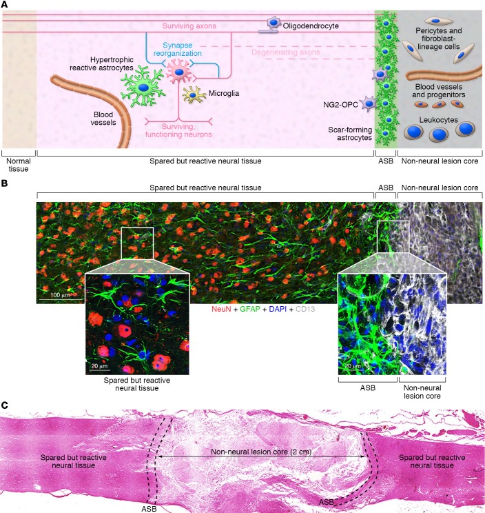
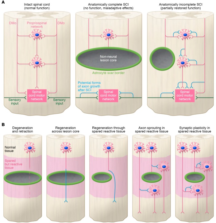
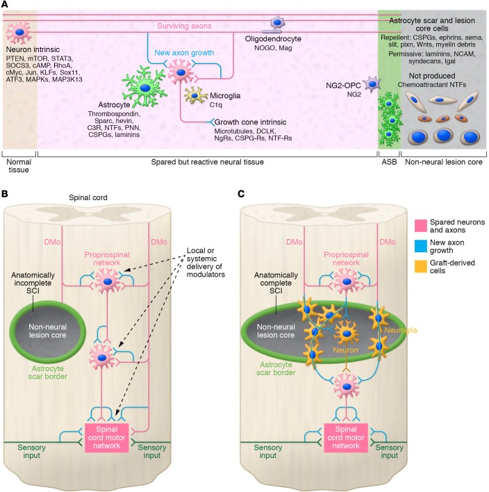
Spinal cord injury (SCI) lesions present diverse challenges for repair strategies. Anatomically complete injuries require restoration of neural connectivity across lesions. Anatomically incomplete injuries may benefit from augmentation of spontaneous circuit reorganization. Here, we review SCI cell biology, which varies considerably across three different lesion-related tissue compartments: (a) non-neural lesion core, (b) astrocyte scar border, and (c) surrounding spared but reactive neural tissue. After SCI, axon growth and circuit reorganization are determined by neuron-cell-autonomous mechanisms and by interactions among neurons, glia, and immune and other cells. These interactions are shaped by both the presence and the absence of growth-modulating molecules, which vary markedly in different lesion compartments. The emerging understanding of how SCI cell biology differs across lesion compartments is fundamental to developing rationally targeted repair strategies.


Conflict of interest statement

Conflict of interest: The authors have declared that no conflict of interest exists.

Figures

Figure 1
Figure 1. SCI lesions exhibit discrete tissue compartments.
(A) Schematic of SCI lesion compartments composed of different neural and non-neural cells. (B) Photomicrograph showing different cellular components in discrete tissue compartments of a mouse severe crush SCI (T9–T10) lesion. Boxed areas are enlarged to show details. Astrocytes are stained green using glial fibrillary acidic protein (GFAP). Neurons are stained red using NeuN. Pericytes and fibroblast-lineage cells in lesion core are stained white using CD13. Cell nuclei are stained blue using DAPI. (C) Survey photomicrograph of human severe SCI (C5–C7) showing relative proportions of lesion compartments (reproduced with permission from Brain, ref. 11). OPC, oligodendrocyte progenitor cell; ASB, astrocyte scar border.
Figure 2
Figure 2. Circuits reorganize after SCI.
(A) Different types of circuit reorganization after different types of SCI. Anatomically complete SCI is associated with synaptic plasticity that can give rise to maladaptive effects such as muscle spasticity or autonomic dysreflexia. Anatomically incomplete SCI can also give rise to axon growth and synaptic plasticity that can partially restore function. (B) Different potential growth responses of axons and synapses after SCI. DMo, descending motor projections.
Figure 3
Figure 3. Growth potential of axons and synapses after SCI can be regulated by diverse cells and molecules in different lesion compartments.
(A) Multiple cell types and molecules can influence and regulate the growth potential of axons and synapses in different lesion compartments. (B) Modulation of axon sprouting, synapse plasticity, or synapse function in spared but reactive and reorganizing neural tissue by delivery of molecules that modify perineuronal net, synapse formation, or synaptic transmission. (C) Transplantation to repopulate non-neural lesion core with neural cells that either provide new relay neurons, or provide neuroglia that support host axon regrowth, to restore circuit connectivity across the lesion. CSPG-Rs, CSPG receptors; KLFs, Krüppel-like factors; NgRs, NOGO-receptor; PNN, Peri-Neuronal-Net; NTFs, neurotrophic factors; NTF-Rs, neurotrophic factor receptors.

Similar articles

Cited by

References

    1. McDonald JW, Sadowsky C. Spinal-cord injury. Lancet. 2002;359(9304):417–425. doi: 10.1016/S0140-6736(02)07603-1. - DOI - PubMed
    1. Anderson KD. Targeting recovery: priorities of the spinal cord-injured population. J Neurotrauma. 2004;21(10):1371–1383. doi: 10.1089/neu.2004.21.1371. - DOI - PubMed
    1. Gupta R, Bathen ME, Smith JS, Levi AD, Bhatia NN, Steward O. Advances in the management of spinal cord injury. J Am Acad Orthop Surg. 2010;18(4):210–222. doi: 10.5435/00124635-201004000-00004. - DOI - PubMed
    1. Barres BA. The mystery and magic of glia: a perspective on their roles in health and disease. Neuron. 2008;60(3):430–440. doi: 10.1016/j.neuron.2008.10.013. - DOI - PubMed
    1. Burda JE, Sofroniew MV. Reactive gliosis and the multicellular response to CNS damage and disease. Neuron. 2014;81(2):229–248. doi: 10.1016/j.neuron.2013.12.034. - DOI - PMC - PubMed