

New Ways to Detect Pediatric Sickle Cell Retinopathy: A Comprehensive Review
- PMID: 28737601
- PMCID: PMC5656515
- DOI: 10.1097/MPH.0000000000000919
New Ways to Detect Pediatric Sickle Cell Retinopathy: A Comprehensive Review
Abstract
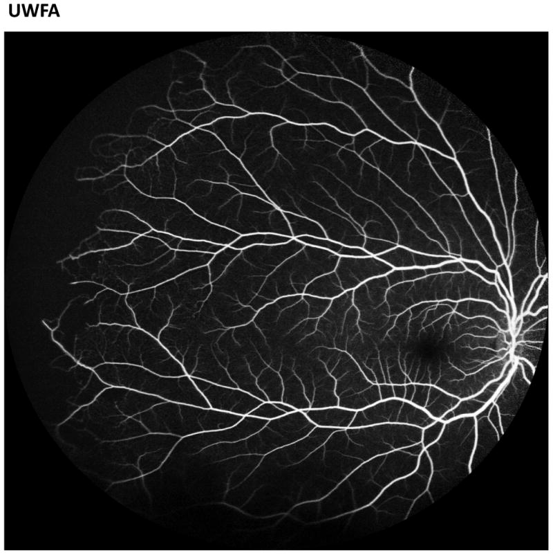
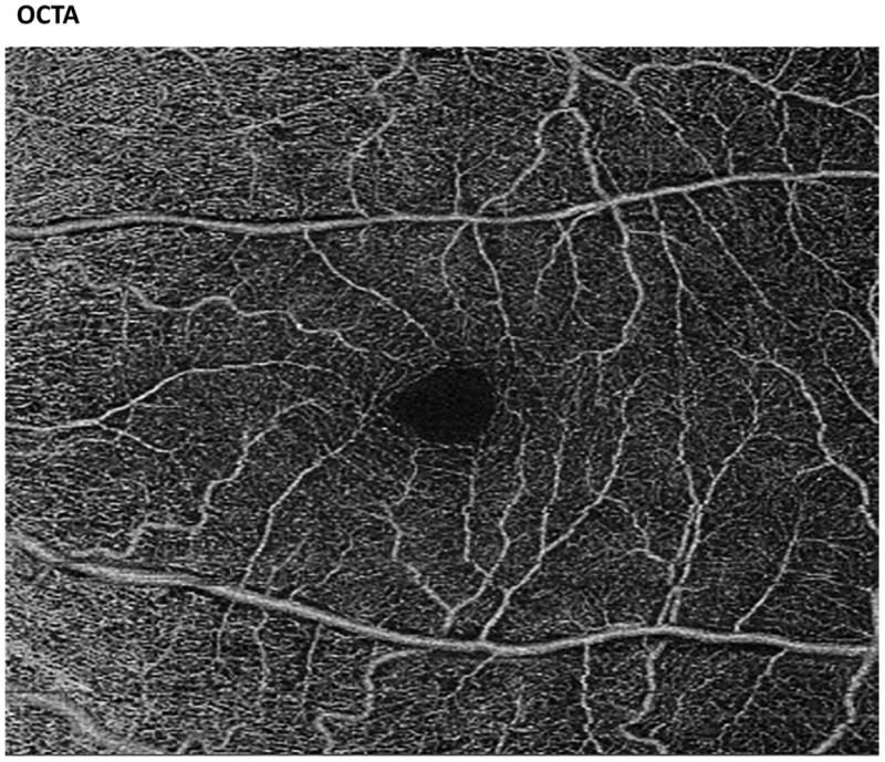
Sickle retinopathy reflects disease-related vascular injury of the eye, which can potentially result in visual loss from vitreous hemorrhage or retinal detachment. Here we review sickle retinopathy among children with sickle cell disease, describe the epidemiology, pediatric risk factors, pathophysiology, ocular findings, and treatment. Newer, more sensitive ophthalmological imaging modalities are available for retinal imaging, including ultra-widefield fluorescein angiography, spectral-domain optical coherence tomography, and optical coherence tomography angiography. Optical coherence tomography angiography provides a noninvasive view of retinal vascular layers that could previously not be imaged and can be quantified for comparative or prospective analyses. Ultra-widefield fluorescein angiography provides a more comprehensive view of the peripheral retina than traditional imaging techniques. Screening for retinopathy by standard fundoscopic imaging modalities detects a prevalence of approximately 10%. In contrast, these more sensitive methods allow for more sensitive examination that includes the retina perimeter where sickle retinopathy is often first detectable. Use of these new imaging modalities may detect a higher prevalence of early sickle pathology among children than has previously been reported. Earlier detection may help in better understanding the pathogenesis of sickle retinopathy and guide future screening and treatment paradigms.
Figures
References
-
- Downes SM, Hambleton IR, Chuang EL, et al. Incidence and natural history of proliferative sickle cell retinopathy: observations from a cohort study. Ophthalmology. 2005;112:1869–1875. - PubMed
-
- Kent D, Arya R, Aclimandos WA, et al. Screening for ophthalmic manifestations of sickle cell disease in the United Kingdom. Eye (Lond) 1994;8(Pt 6):618–622. - PubMed
-
- van Meurs JC. Relationship between peripheral vascular closure and proliferative retinopathy in sickle cell disease. Graefes Arch Clin Exp Ophthalmol. 1991;229:543–548. - PubMed
Publication types
MeSH terms
Grants and funding
LinkOut - more resources
Full Text Sources
Other Literature Sources
Medical
