. 2017 Jul 23;9(7):794.
doi: 10.3390/nu9070794.

Platycodon grandiflorus Root Extract Improves Learning and Memory by Enhancing Synaptogenesis in Mice Hippocampus

Affiliations

Platycodon grandiflorus Root Extract Improves Learning and Memory by Enhancing Synaptogenesis in Mice Hippocampus

Jin-Il Kim et al. Nutrients. .

Abstract

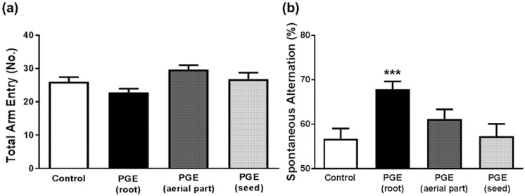
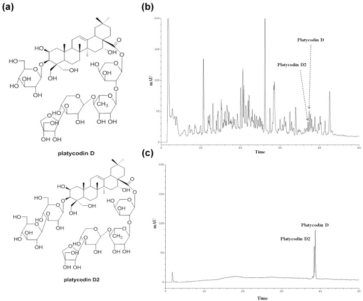
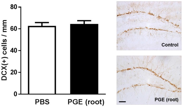
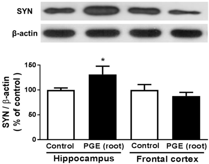
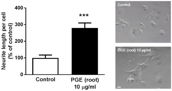
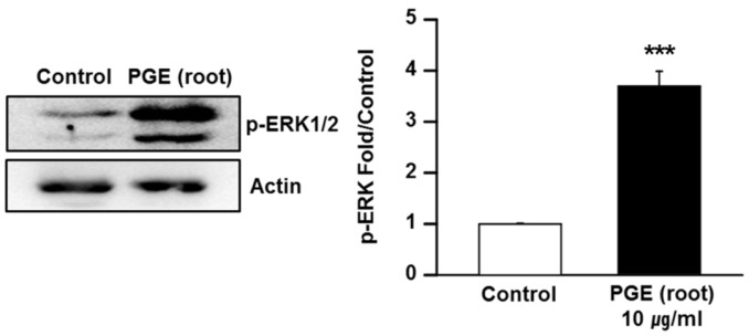
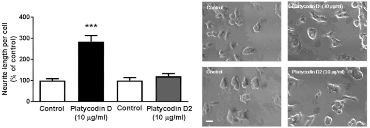
Platycodon grandiflorus (Jacq.) A.DC. (PG) has long been used as an ingredient of foods and is known to have beneficial effects on cognitive functions as well. The present study examined the effect of each PG extract (PGE) from root, aerial part, and seeds on cognitive functions in mice. Changes in spatial learning and memory using a Y-maze test, and markers of adult hippocampal neurogenesis and synaptogenesis were examined. Moreover, changes in neuritogenesis and activation of the ERK1/2 pathway were investigated. Results indicated that mice administered PGE (root) showed increased spontaneous alternation in the Y-maze test and synaptogenesis in the hippocampus. In addition, PGE (root) and platycodin D, the major bioactive compound from the PG root, significantly stimulated neuritic outgrowth by phosphorylation of the ERK1/2 signaling pathway in vitro. These results indicate that the PGE (root), containing platycodin D, enhances cognitive function through synaptogenesis via activation of the ERK1/2 signaling pathway.

Keywords: Platycodon grandiflorus; cognition; neuritogenesis; platycodin D; synaptogenesis.


Conflict of interest statement

The authors declare no conflict of interest.

Figures

Figure 1
Figure 1
Oral administration of PGEs (100 mg/kg/day) for 15 days. PGEs are classified into three different parts including PGE (root), PGE (aerial part), and PGE (seed). The cognition of mice treated with three different PGEs underwent a Y-maze test. After the behavioral experiment, animals administered with PGE (root) were sacrificed and brains were isolated for analysis. Subsequently, immunohistochemistry of brains was performed using DCX antibody and immunoblotting with SYN antibody. PGE, Platycodon grandiflorus extract. DCX, doublecortin. SYN, synaptophysin.
Figure 2
Figure 2
Behavioral test to determine the enhancing effect of PGE on cognition. The Y-maze test shows no significant influence of PGE on total arm entry regardless of the PG structural part administered (a). The PGE (root)-administered group shows a significant increase in spontaneous alternations, indicating PGE (root) significantly increased spatial learning and memory. PGE (aerial part) and PGE (seed)-administered groups show no significant increase of spontaneous alternation (b). Values are the mean + SEM. *** p < 0.05 compared to the vehicle-treated control group. PGE, Platycodon grandiflorus extract.
Figure 3
Figure 3
HPLC chromatograms of the EtOH extract of PG. (a) Structures of platycodin D and platycodin D2, (b) HPLC chromatograms of the root extract, and (c) HPLC chromatograms of standard compounds, platycodin D, and platycodin D2. HPLC analysis was carried out using a gradient elution on a phenomenex Kinetex RP-C18 column (5 µ, 100 A, 150 × 4.6 mm) at a flow rate of 1.0 mL/min, and with UV 210 nm detection.
Figure 4
Figure 4
Immunohistochemical analysis using DCX antibody. DCX is a marker of neurogenesis, especially neuronal fate specification. The PGE (root)-administered group shows a similar number of DCX (+) cells per unit length compared to the control group. Values are the mean + SEM. Scale bar = 50 μm. PGE, Platycodon grandiflorus extract. DCX, doublecortin.
Figure 5
Figure 5
Immunoblotting analysis of synaptophysin expression in the hippocampus and frontal cortex after PGE (root)-administration. The PGE (root)-administered group shows a significantly higher expression of SYN in hippocampus than the controls. The PGE (root)-administered group shows no significant difference in the frontal cortex compared to the controls. Values are the mean + SEM. * p < 0.05 compared to the vehicle-treated control group. PGE, Platycodon grandiflorus extract. SYN, synaptophysin.
Figure 6
Figure 6
In vitro assay to confirm PGE (root)-induced neuritogenesis in PC12 cells. PC12 cells were treated with 10 μg/mL PGE (root) for 24 h and the cells show a significant increase in neurite length compared to the controls. Values are the mean + SEM. *** p < 0.05 compared to the vehicle-treated control group. Scale bar = 20 μm. PGE, Platycodon grandiflorus extract.
Figure 7
Figure 7
Immunoblotting of PGE (root) treated PC12 cells with ERK1/2. Cells were treated with PGE (root) for 24 h and assayed by western blot using anti-ERK1/2 antibodies and anti-phospho-ERK1/2 antibodies (Thr202/Tyr204). PGE-(root) treated PC12 cells show a significant increase in ERK1/2 phosphorylation. Values are the mean + SEM. *** p < 0.05 compared to the vehicle-treated control group. PGE, Platycodon grandiflorus extract.
Figure 8
Figure 8
Measurement of neurite length to confirms the effects of bioactive molecules of PG in PC12 cells. PC12 cells were treated with 10 μg/mL platycodin D or platycodin D2 for 24 h and neurite lengths measured. Platycodin D, but not platycodin D2, treated PC12 cells show significantly increased neurite lengths. Scale bar = 20 μm. Values are the mean + SEM. *** p < 0.05 compared to the vehicle-treated control group.

References

    1. Kondo M., Takei Y., Hirokawa N. Motor protein KIF1A is essential for hippocampal synaptogenesis and learning enhancement in an enriched environment. Neuron. 2012;73:743–757. doi: 10.1016/j.neuron.2011.12.020. - DOI - PubMed
    1. Sun M.K., Hongpaisan J., Lim C.S., Alkon D.L. Bryostatin-1 restores hippocampal synapses and spatial learning and memory in adult fragile x mice. J. Pharmacol. Exp. Ther. 2014;349:393–401. doi: 10.1124/jpet.114.214098. - DOI - PubMed
    1. Liu D., Diorio J., Day J.C., Francis D.D., Meaney M.J. Maternal care, hippocampal synaptogenesis and cognitive development in rats. Nat. Neurosci. 2000;3:799–806. - PubMed
    1. Ramirez-Amaya V., Balderas I., Sandoval J., Escobar M.L., Bermudez-Rattoni F. Spatial long-term memory is related to mossy fiber synaptogenesis. J. Neurosci. 2001;21:7340–7348. - PMC - PubMed
    1. Nordeen K.W., Nordeen E.J. Anatomical and synaptic substrates for avian song learning. J. Neurobiol. 1997;33:532–548. doi: 10.1002/(SICI)1097-4695(19971105)33:5<532::AID-NEU4>3.0.CO;2-5. - DOI - PubMed