. 2017 Sep 7;549(7670):60-65.
doi: 10.1038/nature23479. Epub 2017 Jul 24.

Channel opening and gating mechanism in AMPA-subtype glutamate receptors

Affiliations

Channel opening and gating mechanism in AMPA-subtype glutamate receptors

Edward C Twomey et al. Nature. .

Abstract

AMPA (α-amino-3-hydroxy-5-methyl-4-isoxazole propionic acid)-subtype ionotropic glutamate receptors mediate fast excitatory neurotransmission throughout the central nervous system. Gated by the neurotransmitter glutamate, AMPA receptors are critical for synaptic strength, and dysregulation of AMPA receptor-mediated signalling is linked to numerous neurological diseases. Here we use cryo-electron microscopy to solve the structures of AMPA receptor-auxiliary subunit complexes in the apo, antagonist- and agonist-bound states and determine the iris-like mechanism of ion channel opening. The ion channel selectivity filter is formed by the extended portions of the re-entrant M2 loops, while the helical portions of M2 contribute to extensive hydrophobic interfaces between AMPA receptor subunits in the ion channel. We show how the permeation pathway changes upon channel opening and identify conformational changes throughout the entire AMPA receptor that accompany activation and desensitization. Our findings provide a framework for understanding gating across the family of ionotropic glutamate receptors and the role of AMPA receptors in excitatory neurotransmission.


Conflict of interest statement

The authors declare no competing financial interests.

Figures

Extended Data Figure 1
Extended Data Figure 1. FSC curves for cryo-EM reconstructions
Fourier shell correlation (FSC) curves calculated between half-maps for GluA2-GSG1LZK-1, GluA2-GSG1LZK-2, GluA2-GSG1Lapo-1, GluA2-GSG1Lapo-2 and GluA2-STZGlu+CTZ cryo-EM reconstructions, as well as for the GluA2-STZGlu+CTZ TMD reconstruction from directed refinement. The dashed line indicates FSC=0.143.
Extended Data Figure 2
Extended Data Figure 2. Local resolution and fitting of cryo-EM maps
Local resolution calculated using Resmap and two unfiltered halves of the reconstruction for GluA2-GSG1LZK-1, GluA2-GSG1LZK-2, GluA2-GSG1Lapo-1, GluA2-GSG1Lapo-2 and GluA2-STZGlu+CTZ structures viewed parallel to the membrane as a surface (a, d, h, k, n) and slice through the center of the receptor (b, e, i, l, o), with the cross-validation FSC curves for the refined model versus unfiltered half maps (one used in the refinement, work, and another one not, free) and the unfiltered summed maps shown on the right (c, f, j, m, p).
Extended Data Figure 3
Extended Data Figure 3. Closed state 1 cryo-EM density and comparison of ZK-bound and apo states
a–d, Fragments of GluA2-GSG1LZK-1 and GluA2-GSG1Lapo-1 with the corresponding cryo-EM density: (a,d) ATD and LBD of subunit A in GluA2-GSG1LZK-1 (a) and GluA2-GSG1LApo-1 (d) with density for ZK indicated in the GluA2-2xGSG1LZK-1 structure; (b) M2 helix and (c) selectivity filter with the Q/R-site Q586 side chains pointing towards the center of the pore in GluA2-GSG1LZK-1. e, Superposition of GluA2-GSG1LZK-1 (blue) and GluA2-GSG1Lapo-1 (red) viewed parallel to the membrane. Note, the structures are almost indistinguishable (RMSD = 0.526 Å). Densities are shown at 6 σ.
Extended Data Figure 4
Extended Data Figure 4. Closed state 2 structure and digitonin binding pocket
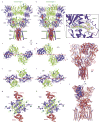
a–b, Structures of GluA2-GSG1LZK-1 (a) and GluA2-GSG1LZK-2 (b) viewed parallel to the membrane. The GluA2 subunits A and C are colored purple, B and D green and GSG1L red. The competitive antagonist ZK and digitonin are shown as space-filling models. In b, inset shows expanded view of the boxed region, demonstrating cryo-EM density for digitonin (blue mesh, 4 σ). Digitonin and the surrounding residues in the inset are shown in stick representation. c–h, Top down views along the axis of the overall 2-fold rotational symmetry on the ATD (c–d), LBD (e–f) and TMD (g–h) layers. Rigid-body rotation of the ATD tetramer in (d) and rotation of LBD dimers in (f) are indicated by red arrows. (i–j) Superposition of GluA2-GSG1LZK-2 (blue) and GluA2-GSG1LApo-2 (red) viewed parallel to the membrane. Note, the structures are almost indistinguishable (RMSD = 0.701 Å).
Extended Data Figure 5
Extended Data Figure 5. Cryo-EM density for the open state
a–f, Fragments of GluA2-STZGlu+CTZ with the corresponding cryo-EM density: (a) zoom on the glutamate binding pocket, (b–c) ion channel pore with a central density at the selectivity filter, likely for a sodium ion that is hydrated based on the pore diameter, viewed (b) from the top of the selectivity filter looking down into the cytoplasm or (c) parallel to the membrane with two distal and proximal GluA2 subunits removed, (d) density for CTZ and (e) transmembrane domain segments for GluA2 (upper row) and STZ (lower row).
Extended Data Figure 6
Extended Data Figure 6. Overview of single-particle cryo-EM and stoichiometry for GluA2-STZ and GluA2-GSG1L solubilized in digitonin
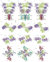
a–b, Two-dimensional class averages for GluA2-STZGlu+CTZ (a) and GluA2-GSG1LZK (b) indicating 3-layer architecture of the particles. c–d, Final densities for GluA2-STZGlu+CTZ (c) and GluA2-GSG1LZK-1 (d) with the GluA2 subunits A and C colored purple, B and D green, STZ cyan and GSG1L red. Insets show 2D slices made parallel to the membrane through the refined, nonfiltered map. Note, while four STZ molecules bind one receptor, only two copies of GSG1L can bind per GluA2 tetramer.
Extended Data Figure 7
Extended Data Figure 7. Conformational differences between the closed, open and desensitized states
a–c, Structures of GluA2-GSG1LZK-1 in the closed state (a), GluA2-STZGlu+CTZ in the open state (b) and GluA2-2xGSG1LQuis in the desensitized state (c) viewed parallel to the membrane. The GluA2 subunits A and C are colored purple, B and D green, GSG1L red and STZ cyan. The competitive antagonist ZK, agonists Glu and Quis and positive allosteric modulator CTZ are shown as space-filling models. (d–l), Top down views along the axis of the overall 2-fold rotational symmetry on the layers of ATD (d–f), LBD (g–i) and TMD (j–l). Rigid-body rotation of the ATD tetramers in (e) and (f), broadening of LBD layer in (h) and rotation of subunit A/C LBDs in (i) are indicated by red arrows. Note, dramatic opening in the middle of the LBD layer (h) and pore dilation (k) in the open state.
Extended Data Figure 8
Extended Data Figure 8. iGluR Gating Mechanism
Two of four iGluR subunits are shown with the ATDs omitted. Four basic states of iGluR gating are illustrated: Resting, represented by apo (GluA2-GSG1Lapo-1) or antagonist-bound closed state (GluA2-GSG1LZK-1) structures; Closed, agonist-bound (pre-active state crystal structures,); Open (GluA2-STZGlu+CTZ) and Desensitized (GluA2-2xGSG1LQuis complex). Transitions between the states are indicated by black arrows, conformational rearrangements by blue arrows and ionic current through the open channel by an orange arrow. Upper and lower gates are indicated by one and two red asterisks, respectively, with red sticks at the upper gate representing channel occluding residues at the bundle crossing and the Q/R site at the lower gate. Glutamate molecules are illustrated by orange wedges. The receptor sits in a resting, closed state, with its LBD clamshells in the maximally open conformations, unoccupied by the neurotransmitter glutamate. Upon glutamate binding, the LBD clamshells close, as described in the pre-activated crystal structures, to an intermediate state that does not put enough strain on the LBD-TMD linkers to open the channel. The LBDs then transition to their maximally-closed state, which strains the LBD-TMD linkers, causing the channel pore to open and conduct ions. Most AMPA receptors, however, quickly desensitize, transitioning to the desensitized state from the open state via the agonist-bound, closed state. Desensitization is accompanied by the rupture of the upper LBD interfaces, with the LBDs adapting their maximally closed clamshell conformations, as described in the desensitized-state GluA2-GSG1L complex.
Figure 1
Figure 1. GluA2-GSG1L and ion channel structure
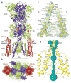
a–b, Model of GluA2-2xGSG1LZK-1 viewed (a) parallel to or (b) from the intracellular side of the membrane with GluA2 subunits A and C colored purple, B and D green and GSG1L red. The competitive antagonists ZK200775 are shown as space-filling models. c, Close-up view of the pore-lining domains M2 and M3 in subunits A and C with cryo-EM density shown as blue mesh. d, Ion conduction pathway (violet) with pore-lining residues in the M2 and M3 segments of subunits A and C shown as sticks.
Figure 2
Figure 2. Cryo-EM of an activated GluA2-STZ complex
a, Representative whole-cell currents recorded at −60 mV membrane potential from a HEK 293 cell expressing GluA2-STZ in response to 1 s applications of Glu alone or in the continuous presence of 30 μM CTZ. b, Two-dimensional class averages of GluA2-STZGlu+CTZ particles, showing diverse orientations. c, 4.2 Å cryo-EM reconstruction of the entire GluA2-STZGlu+STZ complex, with GluA2 subunits colored green and purple, and STZ in blue; viewed perpendicular to the membrane. The dashed outline highlights the area focused on in directed refinement to improve the TMD density. d–e, Density of the GluA2-STZGlu+CTZ TMD at 4.0 Å resolution from directed refinement, viewed parallel (d) and perpendicular (e) to the membrane.
Figure 3
Figure 3. Structure of the GluA2-STZ complex
a–b, Model of GluA2-4xSTZGlu+CTZ viewed parallel to the membrane with GluA2 subunits A and C colored purple, B and D green and STZ cyan. The molecules of agonist Glu and positive allosteric modulator CTZ are shown as space-filling models.
Figure 4
Figure 4. Ion channel pore in open, closed and desensitized states
a, The GluA2-STZGlu+CTZ ion conduction pathway (cyan) with pore-lining residues in M2 and M3 segments of subunits A and C shown as sticks. b, Pore radius calculated using HOLE for GluA2-STZGlu+CTZ in the open state (pink), GluA2-GSG1LZK-1 in the closed state (blue) and GluA2-2xGSG1LQuis in the desensitized state (orange). c–d, TMD ribbon diagrams for the structures in (b) viewed parallel to membrane (c) or extracellularly (d). In (c), subunits B and D as well as M4 segments are removed for clarity.
Figure 5
Figure 5. Structural rearrangements in during gating
GluA2 domains are shown for structures of GluA2-GSG1LZK-1 in the closed state (blue), GluA2-STZGlu+CTZ in the open state (pink) and GluA2-2xGSG1LQuis in the desensitized state (orange). a–d, LBD monomers shown individually (a–c) or in superposition (d). ZK, Glu and Quis molecules are shown in sticks. e–f, Changes in LBD dimer conformation upon transition from closed to open (e) and open to desensitized (f) states. Separation of upper D1 and lower D2 lobes is indicated by arrows. (g–i) LBD tetramers viewed from the ion channel. Broadening of the LBD layer in the open state and rotation of the A and C monomers in the desensitized state are indicated by red arrows. Blue arrows in (i) point to the cleft between the desensitized state LBD protomers signifying the loss of local LBD dimer 2-fold symmetry and 3-fold reduction of intradimer interface. (j–l) S1-M1, M3-S2 and S2-M4 linkers viewed parallel to membrane. The A/C and B/D subunit pairs are shown separately and viewed orthogonally. Distances between Cα atoms of K505, S635 and G771 are indicated. Red and blue stars in (k) indicate the S2-M4 linkers extended towards the pore and one helical turn-unwound M3 helices in A/C subunits and kinked M3 helices in the B/D subunits of the open state, respectively.

References

    1. Kumar J, Mayer ML. Functional insights from glutamate receptor ion channel structures. Annu Rev Physiol. 2013;75:313–337. - PMC - PubMed
    1. Traynelis SF, et al. Glutamate receptor ion channels: structure, regulation, and function. Pharmacol Rev. 2010;62:405–496. - PMC - PubMed
    1. Bowie D. Ionotropic glutamate receptors & CNS disorders. CNS Neurol Disord Drug Targets. 2008;7:129–143. - PMC - PubMed
    1. Chater TE, Goda Y. The role of AMPA receptors in postsynaptic mechanisms of synaptic plasticity. Frontiers in cellular neuroscience. 2014;8:401. - PMC - PubMed
    1. Henley JM, Wilkinson KA. Synaptic AMPA receptor composition in development, plasticity and disease. Nature reviews. Neuroscience. 2016;17:337–350. - PubMed

Publication types

MeSH terms