. 2017 Sep;97(9):1063-1071.
doi: 10.1038/labinvest.2017.64. Epub 2017 Jul 24.

Melanoma subtypes demonstrate distinct PD-L1 expression profiles

Affiliations

Melanoma subtypes demonstrate distinct PD-L1 expression profiles

Genevieve J Kaunitz et al. Lab Invest. 2017 Sep.

Abstract

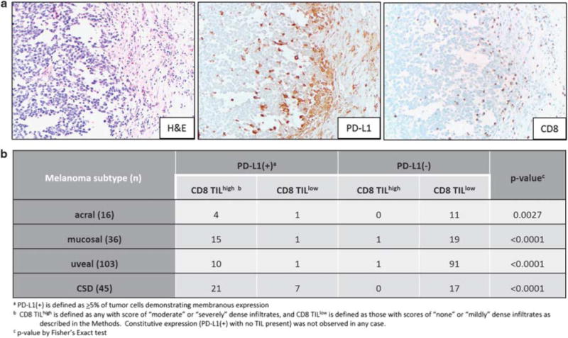
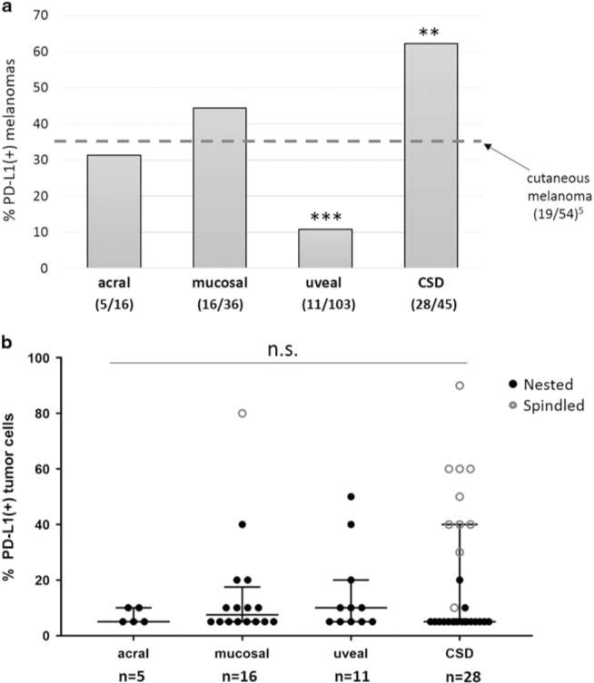
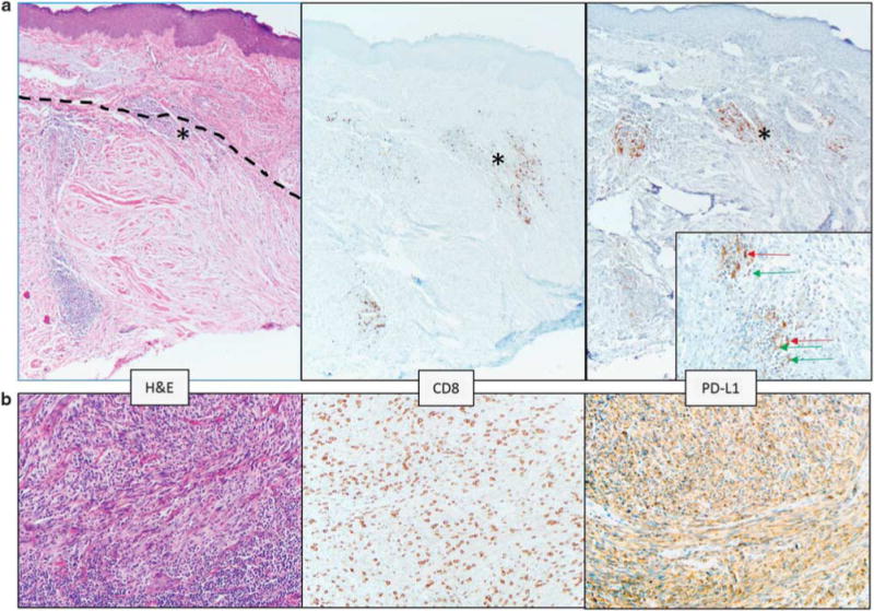
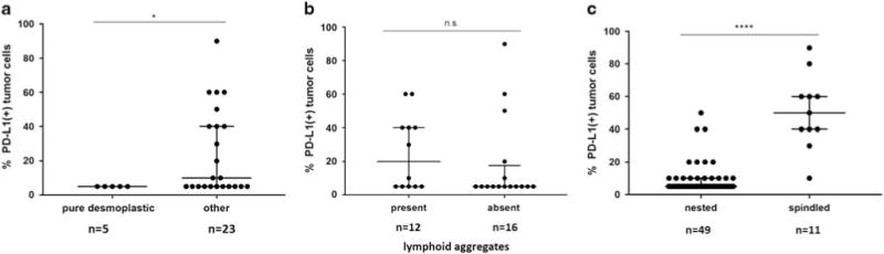
PD-L1 expression in the tumor immune microenvironment is recognized as both a prognostic and predictive biomarker in patients with cutaneous melanoma, a finding closely related to its adaptive (IFN-γ-mediated) mechanism of expression. Approximately 35% of cutaneous melanomas express PD-L1, however, the expression patterns, levels, and prevalence in rarer melanoma subtypes are not well described. We performed immunohistochemistry for PD-L1 and CD8 on 200 formalin-fixed paraffin-embedded specimens from patients with acral (n=16), mucosal (n=36), uveal (n=103), and chronic sun-damaged (CSD) (n=45) melanomas (24 lentigo maligna, 13 'mixed' desmoplastic, and 8 'pure' desmoplastic melanomas). CD8+ tumor-infiltrating lymphocyte (TIL) densities were characterized as mild, moderate, or severe, and their geographic association with PD-L1 expression was evaluated. Discrete lymphoid aggregates, the presence of a spindle cell morphology, and the relationship of these features with PD-L1 expression were assessed. PD-L1 expression was observed in 31% of acral melanomas, 44% of mucosal melanomas, 10% of uveal melanomas, and 62% of CSD melanomas (P<0.0001). Compared to our previously characterized cohort of cutaneous melanomas, the proportion of PD-L1(+) tumors was lower in uveal (P=0.0002) and higher in CSD (P=0.0073) melanomas, while PD-L1 expression in the acral and mucosal subtypes was on par. PD-L1 expression in all subtypes correlated with a moderate-severe grade of CD8+ TIL (all, P<0.003), supporting an adaptive mechanism of expression induced during the host antitumor response. The tumor microenvironments observed in CSD melanomas segregated by whether they were the pure desmoplastic subtype, which showed lower levels of PD-L1 expression when compared to other CSD melanomas (P=0.047). The presence of lymphoid aggregates was not associated with the level of PD-L1 expression, while PD-L1(+) cases with spindle cell morphology demonstrated higher levels of PD-L1 than those with a nested phenotype (P<0.0001). Our findings may underpin the reported clinical response rates for anti-PD-1 monotherapy, which vary by subtype.


Conflict of interest statement

CONFLICT OF INTEREST: The remaining authors declare no conflict of interest.

Figures

Figure 1
Figure 1
All melanoma subtypes demonstrate PD-L1 expression in geographic association with CD8+ tumor-infiltrating lymphocytes (TIL), consistent with an adaptive (IFN-γ-mediated) mechanism of expression. (a) PD-L1 expression on both tumor cells and immune cells is observed at the tumor-host interface in a vulvar mucosal melanoma. 200x original magnification, all panels. (b) The adaptive pattern of expression was consistently observed across all melanoma subtypes studied. There were only two melanomas studied with moderate–severe grade of TIL present that lacked PD-L1 expression (2/54, 4%).
Figure 2
Figure 2
Prevalence and levels of PD-L1 expression vary by histologic subtype of melanoma. (a) Uveal melanomas showed the lowest proportion of cases with PD-L1 expression, while CSD melanomas showed the highest proportion. The proportion of PD-L1(+) tumors in both of these subtypes was significantly different from conventional cutaneous melanoma (***P = 0.0002 and **P = 0.0073, chi-square test), the latter reported previously using the same staining and scoring methods. The proportion of PD-L1(+) cases among acral and mucosal melanomas was not significantly different from that observed in cutaneous disease. (b) PD-L1 expression levels were compared by melanoma subtype in cases that were PD-L1(+). A narrower range of expression levels was observed in acral melanomas when compared to the other subtypes, but significant differences in the median levels of PD-L1 expression were not observed (P = 0.91). n.s., not significant.
Figure 3
Figure 3
Pure desmoplastic melanomas demonstrate a different PD-L1 expression pattern than other CSD melanomas. (a) The dotted line in the left panel delineates the upper boundary of a desmoplastic melanoma centered in the dermis. Solar elastosis is a prominent feature in the superficial dermis. Lymphoid aggregates are present, and one at the boundary of the melanoma and normal dermis is marked with an asterisk (*) and is shown on the inset of the right panel. In the pure desmoplastic melanomas, PD-L1 expression was observed on lymphocytes (green arrow on inset) and macrophages (red arrows on inset) in lymphoid aggregates. This case did not show PD-L1 expression on tumor cells. Original magnification, ×100, inset ×400. (b) CSD melanoma with spindled morphology showing 60% tumor cell PD-L1 expression associated with a ‘severe’ grade CD8+ TIL infiltrate. ‘Severe’ grade is defined as a diffuse infiltrate of TIL throughout the melanoma. ×200 original magnification.
Figure 4
Figure 4
Association of morphologic features with levels of PD-L1 expression. (a) Among PD-L1(+) CSD melanomas, there were significantly higher median PD-L1 expression levels in cases lacking a pure desmoplastic phenotype (*P = 0.047, Mann–Whitney U-test). (b) The presence or absence of lymphoid aggregates did not correlate with the levels of PD-L1 expression observed in PD-L1(+) CSD melanomas (P = 0.22). (c) Among all melanoma subtypes, PD-L1(+) tumors with a spindled morphology demonstrated a higher percentage of tumor cells with PD-L1 display compared to those with a nested morphology (****P<0.0001). n.s., not significant.

References

    1. Sunshine J, Taube JM. PD-1/PD-L1 inhibitors. Curr Opin Pharmacol. 2015;23:32–38. - PMC - PubMed
    1. Daud AI, Wolchok JD, Robert C, et al. Programmed death-ligand 1 expression and response to the anti-programmed death 1 antibody pembrolizumab in melanoma. J Clin Oncol. 2016;34:4102–4109. - PMC - PubMed
    1. Madore J, Vilain RE, Menzies AM, et al. PD-L1 expression in melanoma shows marked heterogeneity within and between patients: implications for anti-PD-1/PD-L1 clinical trials. Pigment Cell Melanoma Res. 2015;28:245–253. - PubMed
    1. Obeid JM, Erdag G, Smolkin ME, et al. PD-L1, PD-L2 and PD-1 expression in metastatic melanoma: Correlation with tumor-infiltrating immune cells and clinical outcome. Oncoimmunology. 2016;5:e1235107. - PMC - PubMed
    1. Taube JM, Anders RA, Young GD, et al. Colocalization of inflammatory response with B7-h1 expression in human melanocytic lesions supports an adaptive resistance mechanism of immune escape. Sci Transl Med. 2012;4:127ra137. - PMC - PubMed

Publication types