. 2017 Jul 24;18(1):560.
doi: 10.1186/s12864-017-3947-4.

Anion-selective Formate/nitrite transporters: taxonomic distribution, phylogenetic analysis and subfamily-specific conservation pattern in prokaryotes

Affiliations

Anion-selective Formate/nitrite transporters: taxonomic distribution, phylogenetic analysis and subfamily-specific conservation pattern in prokaryotes

Mishtu Mukherjee et al. BMC Genomics. .

Abstract

Background: The monovalent anions formate, nitrite and hydrosulphide are main metabolites of bacterial respiration during anaerobic mixed-acid fermentation. When accumulated in the cytoplasm, these anions become cytotoxic. Membrane proteins that selectively transport these monovalent anions across the membrane have been identified and they belong to the family of Formate/Nitrite Transporters (FNTs). Individual members that selectively transport formate, nitrite and hydrosulphide have been investigated. Experimentally determined structures of FNTs indicate that they share the same hourglass helical fold with aquaporins and aquaglyceroporins and have two constriction regions, namely, cytoplasmic slit and central constriction. Members of FNTs are found in bacteria, archaea, fungi and protists. However, no FNT homolog has been identified in mammals. With FNTs as potential drug targets for many bacterial diseases, it is important to understand the mechanism of selectivity and transport across these transporters.

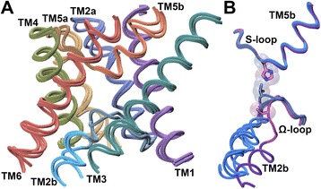
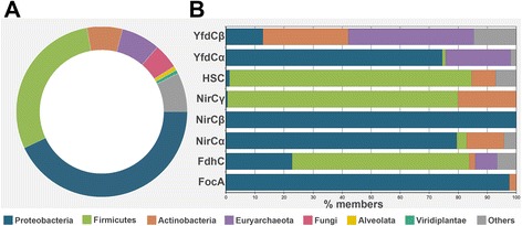
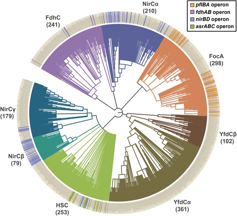
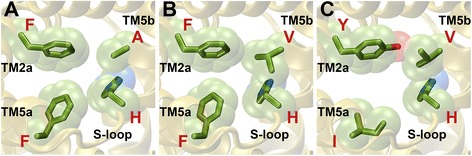
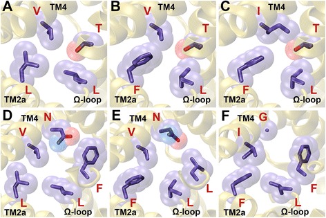
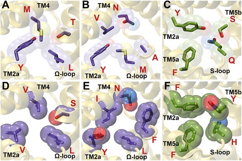
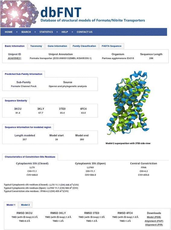
Results: We have systematically searched the sequence databases and identified 2206 FNT sequences from bacteria, archaea and eukaryotes. Although FNT sequences are very diverse, homology modeling followed by structure-based sequence alignment revealed that nearly one third of all the positions within the transmembrane region exhibit high conservation either as a group or at the level of individual residues across all three kingdoms. Phylogenetic analysis of prokaryotic FNT sequences revealed eight different subgroups. Formate, nitrite and hydrosulphide transporters respectively are clustered into two (FocA and FdhC), three (NirC-α, NirC-β and NirC-γ) and one (HSC) subfamilies. We have also recognized two FNT subgroups (YfdC-α and YfdC-β) with unassigned function. Analysis of taxonomic distribution indicates that each subfamily prefers specific taxonomic groups. Structure-based sequence alignment of individual subfamily members revealed that certain positions in the two constriction regions and some residues facing the interior show subfamily-specific conservation. We have also identified examples of FNTs with the two constriction regions formed by residues that are less frequently observed. We have developed dbFNT, a database of FNT models and associated details. dbFNT is freely available to scientific community.

Conclusions: Taxonomic distribution and sequence conservation of FNTs exhibit subfamily-specific features. The conservation pattern in the central constriction and cytoplasmic slit in the open and closed states are distinct for YfdC and NirC subfamilies. The same is true for some residues facing the interior of the transporters. The specific residues in these positions can exert influence on the type of solutes that are transported by these proteins. With FNTs found in many disease-causing bacteria, the knowledge gained in this study can be used in the development and design of anti-bacterial drugs.

Keywords: Anion transport; Channel selectivity and transport; Conformational changes; FNT family; Gating; Homology modeling; Hour-glass helical fold; Phylogenetic analysis; Sequence diversity; Structure-based sequence alignment.


Conflict of interest statement

Ethics approval and consent to participate

Not applicable.

Consent for publication

Not applicable.

Competing interests

Not applicable.

Publisher’s Note

Springer Nature remains neutral with regard to jurisdictional claims in published maps and institutional affiliations.

Figures

Fig. 1
Fig. 1
Superposition of FNT channel structures. (a) Superposition of experimentally determined structures of formate (PDB ID: 3KCU; monomer B;), nitrite (PDB ID: 4FC4; monomer A;) and hydrosulphide (PDB ID: 3TDR; monomer I;) channel structures. Only the backbone of six transmembrane helical segments and the Ω- and S-loops were considered for superposition. Side-chains and the loops connecting the transmembrane segments are not shown for clarity. (b) Superposition of functionally important Ω- and S-loops from five different monomers from three formate channel structures. Monomer A from PDB ID: 3KCU, monomers A and E from PDB ID: 3KLY and monomers A and B from PDB ID: 3Q7K were superposed in the same way as described in Fig. 1A. Only the backbone of the two loop regions is displayed to illustrate the conformational changes of Ω-loop. Molecules plotted in this figure and subsequent figures utilized the VMD software package [72]. Thr-91 from the Ω-loop and His-209 from the S-loop are shown in stick representation. It should be noted that Thr-91 has moved away representing the open state of FNT structure [26]
Fig. 2
Fig. 2
Taxonomic distribution of FNTs. (a) Taxonomic distribution of all 2206 FNT sequences. (b) Taxonomic distribution for each individual prokaryotic FNT subfamily is provided. The species groups to which the channels belong are shown in different colors
Fig. 3
Fig. 3
Phylogenetic analysis of prokaryotic FNTs. Phylogenetic analysis of 1723 prokaryotic FNTs identified eight subgroups. Two subfamilies (FocA and FdhC) for formate channels and three subgroups (NirC-α, NirC-β and NirC-γ) for nitrite channels have been recognized in addition to hydrosulphide (HSC) channels. The uncharacterized YfdC is divided into two clusters (YfdC-α and YfdC-β). The numbers given in the brackets represent the number of FNT channels for each cluster. With 361 and 79 members, Yfdc-α and NirC- β are the largest and smallest clusters respectively. The presence of operon is indicated in the outer circle with the respective color codes. The figure was generated using the web-based tool iTOL (Interactive Tree of Life) [73]
Fig. 4
Fig. 4
Residues showing subfamily-specific variations at the central constriction. Residues of FNT channels forming central constriction are shown for (a) FocA (PDB ID: 3KLY, monomer A), (b) NirC-α (UniProt ID: I1XLF9) and (c) YfdC-α (UniProt ID: U1X6D4). The four residues correspond to Phe-75 (TM2a), Phe-202 (TM5a), His-209 (S-loop) and Ala-212 (TM5b) according to the PDB structure 3KCU numbering. His-209 residue in the S-loop is almost invariant in all the FNT channels. The aromatic character of Phe-75 in TM2a shows near absolute conservation. Ala-212 in TM5b is replaced by Val in NirC-α and YfdC-α subfamilies (Table 4). Phe-202 in TM5a is substituted by other hydrophobic residues such as Ile, Val, Met and Leu in YfdC subfamilies
Fig. 5
Fig. 5
Residues displaying subfamily-specific variation at the cytoplasmic slit. Residues of FNT channels forming the cytoplasmic slit representing the (A, B, C) closed and (D, E, F) open states. The cytoplasmic slit in the closed state is shown for (a) FocA (PDB ID: 3KLY; monomer A), (b) NirC-α (UniProt ID: I1XLF9) and (c) YfdC-α (UniProt ID: U1X6D4). The four residues forming the cytoplasmic slit in the closed state in PDB ID: 3KCU numbering are Leu-79 (TM2a), Leu-89 (Ω-loop), Thr-91 (Ω-loop) and Val-175 (TM4). Leu-89 and Thr-91 are almost invariant across all FNT channels. The hydrophobic character of Val-175 is maintained in all FNT subfamilies. Leu-79 is substituted by an aromatic residue in NirC-α and YfdC subfamilies. The cytoplasmic slit in the open state is formed by Leu-79 (TM2a), Leu-89 (Ω-loop), Phe-90 (Ω-loop), Asn-172 (TM4) and Val-175 (TM4) and is shown for (d) FocA (PDB ID: 3KLY; monomer E), (e) NirC-α (UniProt ID: I1XLF9) and (f) YfdC-α (UniProt ID: U1X6D4). Open states for NirC-α and YfdC-α were modeled similar to the procedure to model the open states of FocA channel described in the Methods section. Phe-90 shows absolute conservation in FocA subfamily and the aromatic character is nearly 100% conserved in YfdC-α subfamily. However, Asn-172, while showing absolute conservation in FocA and NirC-α subfamilies, is replaced by a Gly residue in YfdC subfamilies
Fig. 6
Fig. 6
FNT channels with unique constriction sites. FNT channels with less hydrophobic cytoplasmic slit in the (a) closed (UniProt ID: L8XVV6 from YfdC-α; CHI value: +3.7) and (b) open (UniProt ID: D5BVC1 from NirC-α; CHI value: +3.1) states. (c) FNT channel with less hydrophobic central constriction site (UniProt ID: Q1Q4Z9- unclassified; CHI value: -2.8). (d) FNT channel with a wider cytoplasmic slit in the closed state (UniProt ID: M0BPP6 from YfdC-β; CVV value: 407 Ǻ3). (e) FNT channel with a narrower cytoplasmic slit in the open state (UniProt ID: I0JHL8 from an unclassified FNT channel; CVV value: 620 Ǻ3). (f) FNT channel with a narrower central constriction (UniProt ID: B9DL87 from NirC-γ; CVV value: 529 Ǻ3)
Fig. 7
Fig. 7
A sample record from dbFNT database for a member of FNT channel family. Screenshot showing the sample record for a FNT channel. In addition to details about FNT subfamily, its source and operon information (if available), the residues forming the two constriction regions along with their CHI and CVV values are also provided. Details about the multiple templates and the RMSD (in Ǻ) values of the model with respect to each template are available. The user can download the sequence and the modeled structures in FASTA and PDB formats respectively

References

    1. Clegg S, Yu F, Griffiths L, Cole JA. The roles of the polytotic membrane proteins NarK, NarU and NirC in Escherichia Coli K-12: two nitrate and three nitrite transporters. Mol Microbiol. 2002;44:143–55. doi: 10.1046/j.1365-2958.2002.02858.x. - DOI - PubMed
    1. Kabil O, Banerjee R. Redox biochemistry of hydrogen sulfide. J Biol Chem. 2010;285:21903–21907. doi: 10.1074/jbc.R110.128363. - DOI - PMC - PubMed
    1. Oren A. Anaerobic respiration. Chichester: John Wiley & Sons Ltd.; 2009.
    1. Stokes JL. Fermentation of glucose by suspensions of Escherichia Coli. J Bacteriol. 1949;57:147–158. - PMC - PubMed
    1. Sawers RG. Differential turnover of the multiple processed transcripts of the Escherichia Coli focA-pflB operon. Microbiology. 2006;152:2197–2205. doi: 10.1099/mic.0.28951-0. - DOI - PubMed

Publication types

LinkOut - more resources